Przejdź do zawartości
Merck
Wszystkie zdjęcia(7)

Key Documents

MAB377

Sigma-Aldrich

Anti-NeuN Antibody, clone A60

clone A60, Chemicon®, from mouse

Synonim(y):

Neuron-Specific Nuclear Protein, Neuna60, A60

Zaloguj sięWyświetlanie cen organizacyjnych i kontraktowych


About This Item

Kod UNSPSC:
12352203
eCl@ss:
32160702
NACRES:
NA.41

pochodzenie biologiczne

mouse

Poziom jakości

forma przeciwciała

purified immunoglobulin

rodzaj przeciwciała

primary antibodies

klon

A60, monoclonal

reaktywność gatunkowa

avian, pig, chicken, human, rat, salamander, ferret, mouse

producent / nazwa handlowa

Chemicon®

metody

flow cytometry: suitable
immunocytochemistry: suitable
immunofluorescence: suitable
immunohistochemistry (formalin-fixed, paraffin-embedded sections): suitable
immunoprecipitation (IP): suitable
western blot: suitable

izotyp

IgG1

Warunki transportu

wet ice

docelowa modyfikacja potranslacyjna

unmodified

informacje o genach

Opis ogólny

Anti-NeuN antibody (NEUronal Nuclei; clone A60) specifically recognizes the DNA-binding, neuron-specific protein NeuN, which is present in most CNS and PNS neuronal cell types of all vertebrates tested. NeuN protein distributions are apparently restricted to neuronal nuclei, perikarya and some proximal neuronal processes in both fetal and adult brain although, some neurons fail to be recognized by NeuN at all ages: INL retinal cells, Cajal-Retzius cells, Purkinje cells, inferior olivary and dentate nucleus neurons, and sympathetic ganglion cells are examples (Mullen et al., 1992; Wolf et al., 1996). Immunohistochemically detectable NeuN protein first appears at developmental timepoints that correspond with the withdrawal of the neuron from the cell cycle and/or with the initiation of terminal differentiation of the neuron (Mullen et al., 1992). Immunoreactivity appears around E9.5 in the mouse neural tube and is extensive throughout the developing nervous system by E12.5. Strong nuclear staining suggests a nuclear regulatory protein function; however, no evidence currently exists as to whether the NeuN protein antigen has a function in the distal cytoplasm or whether it is merely synthesized there before being transported back into the nucleus. No difference between protein isolated from purified nuclei and whole brain extract on immunoblots has been found (Mullen et al., 1992).

Specyficzność

MILLIPORE′s exclusive monoclonal antibody to vertebrate neuron-specific nuclear protein called NeuN (or Neuronal Nuclei) reacts with most neuronal cell types throughout the nervous system of mice including cerebellum, cerebral cortex, hippocampus, thalamus, spinal cord and neurons in the peripheral nervous system including dorsal root ganglia, sympathetic chain ganglia and enteric ganglia. Developmentally, immunoreactivity is first observed shortly after neurons have become postmitotic, no staining has been observed in proliferative zones. The immunohistochemical staining is primarily localized in the nucleus of the neurons with lighter staining in the cytoplasm. The few cell types not reactive with MAB377 include Purkinje, mitral and photoreceptor cells. The antibody is an excellent marker for neurons in primary cultures and in retinoic acid-stimulated P19 cells. It is also useful for identifying neurons in transplants.

Immunogen

Purified cell nuclei from mouse brain

Zastosowanie

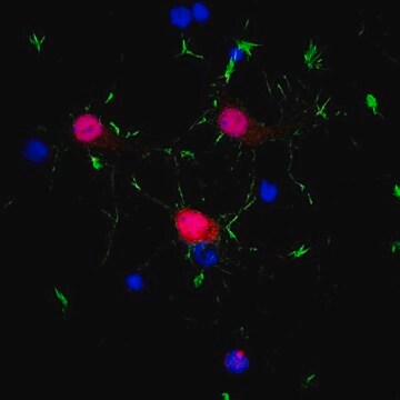
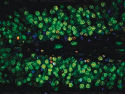
Anti-NeuN Antibody, clone A60 detects level of NeuN and has been published and validated for use in FC, IC, IF, IH, IH(P), IP and WB.
Research Category
Neuroscience
Research Sub Category
Neuronal & Glial Markers
Western Blot Analysis:
A previous lot of this antibody recognized 2-3 bands in the 46-48 kDa range and possibly another band at approximately 66 kDa.

Immunocytochemistry:
1:10-1:100 dilution from a previous lot was used. Neurons in culture should be permeablized with 0.1% triton X-100. All primary antibody dilutions should be performed with simple solutions containing only buffer and primary antibody without excess protein blocks or detergents.

Immunohistochemistry:
1:100-1:1,000. The antibody works best on polyester wax embedded tissue but also works on paraffin embedded tissue at a lower working dilution. The antibody works well with formaldehyde-based fixatives. Citric acid and microwave pretreatment has been used successfully (Sarnat, 1998).

Immunohistochemistry(paraffin) Analysis: A previous lot was used for IH(P).

Optimal working dilutions must be determined by end user.

Jakość

Routinely evaluated by immunohistochemistry on brain tissue.

Immunohistochemistry(paraffin) Analysis:
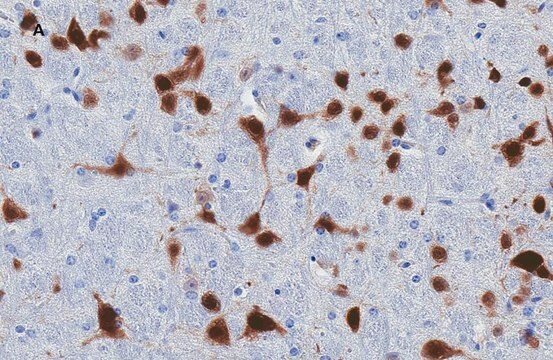
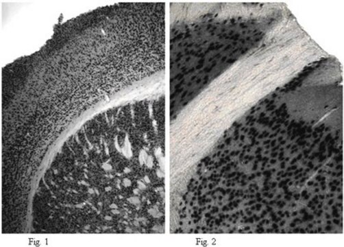
NeuN (cat. # MAB377) staining pattern/morphology in rat cerebellum. Tissue pretreated with Citrate, pH 6.0. This lot of antibody was diluted to 1:100, using IHC-Select Detection with HRP-DAB. Immunoreactivity is seen as nuclear staining in the neurons in the granular layer. Note that there is no signal detected in the nucleus of Purkinje cells.
Optimal Staining With Citrate Buffer, pH 6.0, Epitope Retrieval: Rat Cerebellum

Opis wartości docelowych

46/48 kDa

Postać fizyczna

Format: Purified
Protein A purified
Purified mouse immunoglobulin IgG1 liquid in buffer containing 0.02 M phosphate buffer, 0.25 M NaCl, pH 7.6 with 0.1% sodium azide.

Przechowywanie i stabilność

Stable for 6 months at 2-8ºC from date of receipt.

Komentarz do analizy

Control
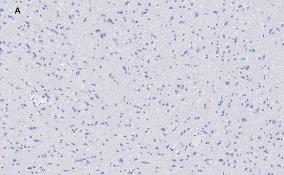
Positive control -Brain Tissue. Negative control - Any non neuronal tissue eg Fibroblasts

Informacje prawne

CHEMICON is a registered trademark of Merck KGaA, Darmstadt, Germany

Oświadczenie o zrzeczeniu się odpowiedzialności

Unless otherwise stated in our catalog or other company documentation accompanying the product(s), our products are intended for research use only and are not to be used for any other purpose, which includes but is not limited to, unauthorized commercial uses, in vitro diagnostic uses, ex vivo or in vivo therapeutic uses or any type of consumption or application to humans or animals.
This page may contain text that has been machine translated.

Nie możesz znaleźć właściwego produktu?  

Wypróbuj nasz Narzędzie selektora produktów.

Kod klasy składowania

12 - Non Combustible Liquids

Klasa zagrożenia wodnego (WGK)

WGK 2

Temperatura zapłonu (°F)

Not applicable

Temperatura zapłonu (°C)

Not applicable


Certyfikaty analizy (CoA)

Poszukaj Certyfikaty analizy (CoA), wpisując numer partii/serii produktów. Numery serii i partii można znaleźć na etykiecie produktu po słowach „seria” lub „partia”.

Masz już ten produkt?

Dokumenty związane z niedawno zakupionymi produktami zostały zamieszczone w Bibliotece dokumentów.

Odwiedź Bibliotekę dokumentów

Presubiculum stimulation in vivo evokes distinct oscillations in superficial and deep entorhinal cortex layers in chronic epileptic rats.
Tolner, EA; Kloosterman, F; van Vliet, EA; Witter, MP; Silva, FH; Gorter, JA
The Journal of Neuroscience null
A U Zaidi et al.
The journal of histochemistry and cytochemistry : official journal of the Histochemistry Society, 48(10), 1369-1375 (2000-09-16)
To understand the biological relationships among various molecules, it is necessary to define the cellular expression patterns of multiple genes and gene products. Relatively simple methods for performing multi-label immunohistochemical detection are available. However, there is a paucity of techniques
Isotropic fractionator: a simple, rapid method for the quantification of total cell and neuron numbers in the brain.
Herculano-Houzel, Suzana and Lent, Roberto
The Journal of Neuroscience, 25, 2518-2521 (2005)
A role for prefrontal cortex in memory storage for trace fear conditioning.
Runyan, Jason D, et al.
The Journal of Neuroscience, 24, 1288-1295 (2004)
Different expression patterns of Bcl-2, Bcl-xl, and Bax proteins after sublethal forebrain ischemia in C57Black/Crj6 mouse striatum.
Wu, C; Fujihara, H; Yao, J; Qi, S; Li, H; Shimoji, K; Baba, H
Stroke null

Produkty

Przeciwciała łączą się z określonymi antygenami w celu wytworzenia ekskluzywnego kompleksu przeciwciało-antygen. Dowiedz się więcej o naturze tego wiązania i jego wykorzystaniu jako znacznika molekularnego w badaniach.

Poznaj różnice między przeciwciałami monoklonalnymi i poliklonalnymi, w tym sposób generowania przeciwciał, numery klonów i formaty przeciwciał.

Immunofluorescencja wykorzystuje cząsteczki fluorescencyjne sprzężone z przeciwciałami do lokalizacji białek, potwierdzania modyfikacji i wizualizacji kompleksów białkowych.

Końcówki do wyboru barwników do cytometrii przepływowej dopasowują fluorofory do konfiguracji cytometru przepływowego, zwiększając wydajność panelu.

Protokoły

Zapoznaj się z naszym przewodnikiem po cytometrii przepływowej, aby odkryć podstawy cytometrii przepływowej, tradycyjne komponenty cytometru przepływowego, kluczowe etapy protokołu cytometrii przepływowej i odpowiednie kontrole.

Nasz zespół naukowców ma doświadczenie we wszystkich obszarach badań, w tym w naukach przyrodniczych, materiałoznawstwie, syntezie chemicznej, chromatografii, analityce i wielu innych dziedzinach.

Skontaktuj się z zespołem ds. pomocy technicznej