おすすめの製品
詳細
Actin, alpha skeletal muscle (UniProt P68135; also known as Alpha-actin-1) is encoded by the ACTA1 (also known as ACTA) gene (Gene ID 100009506) in Oryctolagus cuniculus (Rabbit) species. Actins exist in a variety of structural states, depending on the specific ionic conditions or the interaction with ligand proteins. The oligomeric and polymeric forms that actin molecules assume are dependent on the distinct conformations they adopt. Research indicates distinct actin conformations/structures in the nucleus and the cytoplasm of different cell types and that their distribution varies in response to external signals. In additon to serving as the basic building blocks of cytoskeletal microfilaments, actins are involved in multiple nuclear functions and adopt specific conformations that are important for their association with nuclear actin-binding proteins.
特異性
Clone 2G2 recognizes a tripartite epitope identified in a region of the actin monomer (G-actin) that is not exposed on the surface of filamentous F-actin, but is accessible on the surface of profilin-complexed actin molecule (Gonsior, S.M., et al. (1999). J. Cell Sci. 112 (Pt 6):797-809).
免疫原
Epitope: Nonsequential epitope formed by regions within a.a. 104-226.
Reconstituted complex of rabbit skeletal muscle actin and calf thymus profilin.
アプリケーション
Research Category
細胞骨格
細胞骨格
Anti-Actin Antibody, clone 2G2 is an antibody against Actin for use in Immunocytochemistry, Western Blotting.
Immunocytochemistry Analysis: 5.0 µg/mL from a representative lot detected Actin in serum-starved C2C12 mouse myoblasts.
Immunocytochemistry Analysis: A representative lot showed fixation-dependent cellular staining by fluorescent immunocytochemistry. Clone 2G2 stained nuclear dot-like structures, but not cytoplasmic myofibrils and stress-fibers (MF & SF) in formaldehyde-fixed chicken and mouse myogenic cells, while MF & SF, but not nuclei staining was seen in methanol-fixed chicken cardiac myocytes, fibroblasts, and in vitro differentiated chicken skeletal muscle myotubes (Gonsior, S.M., et al. (1999). J. Cell Sci. 112 (Pt 6):797-809).
Immunocytochemistry Analysis: A representative lot detected nuclear actin immunoreactivity in formaldehyde-fixed COS and NIH-3T3 fibrobastic cells, normal rat kidney NRK epithelial cells, and Xenopus oocytes by fluorescent immunocytochemistry (Gonsior, S.M., et al. (1999). J. Cell Sci. 112 (Pt 6):797-809).
Immunocytochemistry Analysis: A representative lot, when microjected into FH12 rat fibroblasts prior to formaldehyde fixation, showed dot-like staining of cytoplasmic actin monomer (G-actin), but not the microfilament (e.g. myofibrils and stress-fibers) structures (F-actin) seen with FITC-phalloidin staining (Gonsior, S.M., et al. (1999). J. Cell Sci. 112 (Pt 6):797-809).
Immunocytochemistry Analysis: A representative lot detected subnuclear compartmentalized ss-actin immunoreactive foci in onion (Allium cepa) cells by immunofluorescent confocal microscopy (Cruz, J.R., et al. (2009). Chromosoma. 118(2):193-207).
Immunocytochemistry Analysis: A representative lot detected actin immunoreactivity in the nucleus, in the submembraneous actin web, and at the filopodia tips by fluorescent immunocytochemistry staining of fixed rat fibroblasts and HeLa cells (Jockusch, B.M., et al. (2006). Trends Cell Biol. 16(8):391-396).
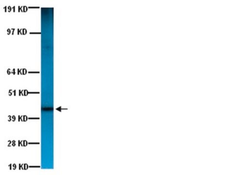
Western Blotting Analysis: A representative lot showed a broad-spectrum species (rabbit, chicken, mouse) reactivity against SDS-denatured actins (alpha, beta and gamma) by Western blotting. However, clone 2G2 displayed no reactivity toward slime mold (Physarum polycephalum) actin or actins from muscle tissues of several invertebrate species (Gonsior, S.M., et al. (1999). J. Cell Sci. 112 (Pt 6):797-809).
Western Blotting Analysis: A representative lot detected actin in all soluble and insoluble fractions of onion (Allium cepa) nuclei preparations (Cruz, J.R., et al. (2009). Chromosoma. 118(2):193-207).
Western Blotting Analysis: A representative lot detected an internal actin fragment corresponding to a.a. 104-226 of rabbit skeletal muscle alpha-actin obtained by limited proteolytic digestion (Gonsior, S.M., et al. (1999). J. Cell Sci. 112 (Pt 6):797-809).
Research Sub Category
Cytoskeleton
Cytoskeleton
品質
Evaluated by Immunocytochemistry in serum-starved HeLa cells.
Immunocytochemistry Analysis: 5.0 µg/mL of this antibody detected Actin in serum-starved HeLa cells.
Immunocytochemistry Analysis: 5.0 µg/mL of this antibody detected Actin in serum-starved HeLa cells.
ターゲットの説明
~42 kDa calculated
物理的形状
Format: Purified
Purified mouse monoclonal IgMκ antibody in PBS with 0.05% sodium azide.
保管および安定性
Stable for 1 year at 2-8°C from date of receipt.
その他情報
Concentration: Please refer to lot specific datasheet.
免責事項
Unless otherwise stated in our catalog or other company documentation accompanying the product(s), our products are intended for research use only and are not to be used for any other purpose, which includes but is not limited to, unauthorized commercial uses, in vitro diagnostic uses, ex vivo or in vivo therapeutic uses or any type of consumption or application to humans or animals.
適切な製品が見つかりませんか。
製品選択ツール.をお試しください
保管分類コード
12 - Non Combustible Liquids
WGK
WGK 2
引火点(°F)
Not applicable
引火点(℃)
Not applicable
適用法令
試験研究用途を考慮した関連法令を主に挙げております。化学物質以外については、一部の情報のみ提供しています。 製品を安全かつ合法的に使用することは、使用者の義務です。最新情報により修正される場合があります。WEBの反映には時間を要することがあるため、適宜SDSをご参照ください。
Jan Code
MABT826:
試験成績書(COA)
製品のロット番号・バッチ番号を入力して、試験成績書(COA) を検索できます。ロット番号・バッチ番号は、製品ラベルに「Lot」または「Batch」に続いて記載されています。
Katja Burk et al.
Scientific reports, 7(1), 2149-2149 (2017-05-21)
The sorting of activated receptors into distinct endosomal compartments is essential to activate specific signaling cascades and cellular events including growth and survival. However, the proteins involved in this sorting are not well understood. We discovered a novel role of
ライフサイエンス、有機合成、材料科学、クロマトグラフィー、分析など、あらゆる分野の研究に経験のあるメンバーがおります。.
製品に関するお問い合わせはこちら(テクニカルサービス)