Pular para o conteúdo
Merck
Todas as fotos(6)

Documentos Principais

M8421

Sigma-Aldrich

Monoclonal Anti-Myosin (Skeletal, Slow) antibody produced in mouse

clone NOQ7.5.4D, ascites fluid

Sinônimo(s):

Anti-CMD1S, Anti-CMH1, Anti-MPD1, Anti-MYHCB, Anti-SPMD, Anti-SPMM

Faça loginpara ver os preços organizacionais e de contrato


About This Item

Número MDL:
Código UNSPSC:
12352203
NACRES:
NA.41

fonte biológica

mouse

Nível de qualidade

conjugado

unconjugated

forma do anticorpo

ascites fluid

tipo de produto de anticorpo

primary antibodies

clone

NOQ7.5.4D, monoclonal

contém

15 mM sodium azide

reatividade de espécies

sheep, rat, bovine, hamster, pig, canine, feline, goat, chicken, mouse, rabbit, human, guinea pig

embalagem

antibody small pack of 25 μL

técnica(s)

electron microscopy: suitable
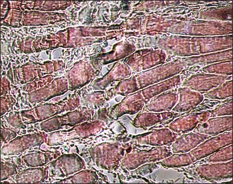
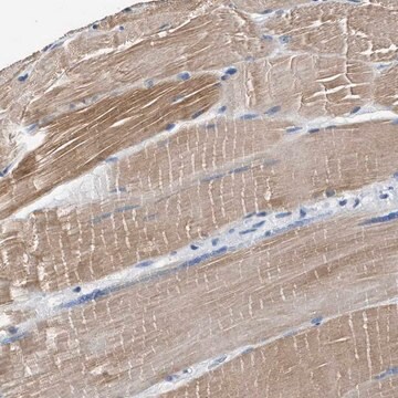
immunohistochemistry (formalin-fixed, paraffin-embedded sections): 1:4,000 using protease-digested, sections of rabbit tongue
indirect ELISA: suitable
radioimmunoassay: suitable
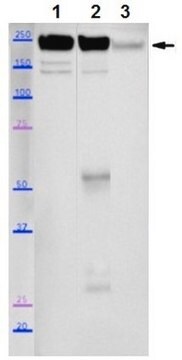
western blot: 1:5,000 using extract of rat or rabbit tongue

Isotipo

IgG1

nº de adesão UniProt

Condições de expedição

dry ice

temperatura de armazenamento

−20°C

modificação pós-traducional do alvo

unmodified

Informações sobre genes

human ... MYH7(4625)
mouse ... Myh7(140781)
rat ... Myh7(29557)

Descrição geral

Myosin is a 480 kDa protein known to interact with actin in muscle and in non-muscle cells. It contains two identical heavy chains (200 kDa each) and four light chains (15-26 kDa). Myosin 7 (MYH7) is mapped to human chromosome 14q11.1.
Monoclonal Anti-Myosin (Skeletal, Slow) (mouse IgG1 isotype) is derived from the NOQ7.5.4D hybridoma produced by the fusion of mouse myeloma cells and splenocytes from BALB/c mice. Myosin, purified from myofibrils isolated from human skeletal muscle, was used as the immunogen.1-3. The isotype is determined by a double diffusion immunoassay using Mouse Monoclonal Antibody Isotyping Reagents, Catalog Number ISO2.

Especificidade

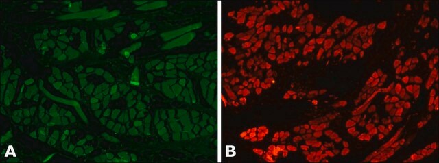
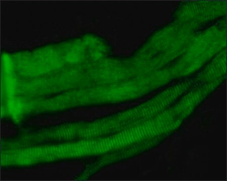
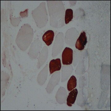
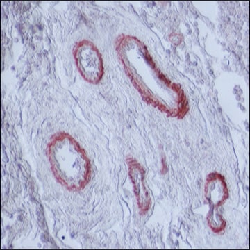
Monoclonal Anti-Myosin (Skeletal, Slow) recognizes an epitope located on the heavy meromyosin portion of human adult skeletal muscle slow myosin. Cross-reactivity has been observed with human, bovine, sheep, goat, porcine, dog, cat, rabbit, hamster, guinea pig, rat, mouse, and chicken. In rat, the product is highly specific for the slow myosin heavy chain of adult skeletal muscle. It does not react with the fast myosin heavy chain. It does not cross-react with embryonic or neonatal rat skeletal myosins, nor with cat fetal/embryonic skeletal myosin. Using immunohistochemical staining, the antibody reacts with human adult type I skeletal muscle fibers and with cardiac muscle cells. It also reacts faintly with adult human C-Fibers, co-expressing myosin heavy chain I and IIa in variable ratios. The product shows no reactivity with human or rat adult smooth muscle or with non-muscle tissues.
Reacts with human adult skeletal muscle slow myosin. Does not react with the fast myosin heavy chain nor with embryonic or neonatal rat skeletal myosins, nor with cat fetal/embryonic skeletal myosin. Also reacts with human adult type I skeletal muscle fibers and with cardiac muscle cells. No reactivity detected with human or rat adult smooth muscle or with non-muscle tissues.

Imunogênio

Human skeletal muscle myosin purified from myofibrils.

Aplicação

Monoclonal Anti-Myosin (Skeletal, Slow) may be used for the localization of slow (Type I) myosin.
Monoclonal Anti-Myosin (Skeletal, Slow) antibody has been used in the detection of Myosin 7 using:
  • light microscopy
  • immunofluorescence staining
  • immunoblotting
  • ELISA
  • solid-phase RIA
  • immunohistology (frozen, formalin-fixed, paraffin-embedded and methacarn-fixed paraffin-embedded tissue sections)
  • immunoelectronmicroscopy
Detection of slow myosin in formalin-fixed, paraffin-embedded, tissues is greatly enhanced by proteolytic digestion of the preparation.

Ações bioquímicas/fisiológicas

Myosin molecules consist of two major regions: tail (rod) and heads; they aggregate into filaments through the tail region and interact with actin and with ATP through the head region. Multiple forms of myosin heavy chains exist for each muscle type: skeletal, cardiac, smooth and in non-muscle. Myosin isoforms exist in different types of skeletal muscle, depending on the physiological function of the muscle. Mammalian muscle fibers are classified primarily into slow (I), fast-red (IIa) and fast-white (IIb) major types. Changes in the speed of muscle contraction brought about by neural influences result from changes in the pattern of expression of myosin and other myofibrillar genes.
Transient expression of different myosin isoforms occurs during fetal growth and development. Mutations in myosin 7 is associated with laing distal myopathy (LDM). Myosin 7 gene mutations results in muscular dystrophy diseases like scapuloperoneal myopathy. Mutations leads to storage of myosin protein aggregates in muscle, leading to myosin storage myopathy. Mutations in MYH7 is also associated with hypertrophic cardiomyopathy and in heart malformation disease called ebstein anomaly.

forma física

Supplied as ascites fluid with 15 mM sodium azide as a preservative.

Armazenamento e estabilidade

For continuous use, store at 2-8 °C for up to one month. For extended storage freeze in working aliquots. Repeated freezing and thawing, or storage in "frostfree" freezers, is not recommended. If slight turbidity occurs upon prolonged storage, clarify the solution by centrifugation before use.

Exoneração de responsabilidade

Unless otherwise stated in our catalog or other company documentation accompanying the product(s), our products are intended for research use only and are not to be used for any other purpose, which includes but is not limited to, unauthorized commercial uses, in vitro diagnostic uses, ex vivo or in vivo therapeutic uses or any type of consumption or application to humans or animals.

Não está encontrando o produto certo?  

Experimente o nosso Ferramenta de seleção de produtos.

Código de classe de armazenamento

12 - Non Combustible Liquids

Classe de risco de água (WGK)

WGK 3

Ponto de fulgor (°F)

Not applicable

Ponto de fulgor (°C)

Not applicable


Escolha uma das versões mais recentes:

Certificados de análise (COA)

Lot/Batch Number

Não está vendo a versão correta?

Se precisar de uma versão específica, você pode procurar um certificado específico pelo número do lote ou da remessa.

Já possui este produto?

Encontre a documentação dos produtos que você adquiriu recentemente na biblioteca de documentos.

Visite a Biblioteca de Documentos

TGFβ contributes to impaired exercise response by suppression of mitochondrial key regulators in skeletal muscle.
Bohm A, et al.
Diabetes, db151723-db151723 (2016)
Developmental myosins: expression patterns and functional significance.
Schiaffino S, et al.
Skeletal Muscle, 5(1), 22-22 (2015)
A de novo mutation of the MYH7 gene in a large Chinese family with autosomal dominant myopathy.
Oda T, et al.
Human Genome Variation, 2, 15022-15022 (2015)
Mutations in the sarcomere gene MYH7 in Ebstein anomaly.
Postma AV, et al.
Circulation: Genomic and Precision Medicine, 4(1), 43-50 (2011)
The hypertrophic cardiomyopathy myosin mutation R453C alters ATP binding and hydrolysis of human cardiac β-myosin.
Bloemink M, et al.
The Journal of Biological Chemistry, 289(8), 5158-5167 (2014)

Nossa equipe de cientistas tem experiência em todas as áreas de pesquisa, incluindo Life Sciences, ciência de materiais, síntese química, cromatografia, química analítica e muitas outras.

Entre em contato com a assistência técnica