Wszystkie zdjęcia(1)
Kluczowe dokumenty
Z764000
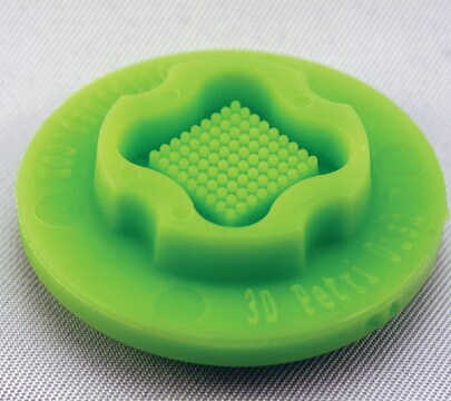
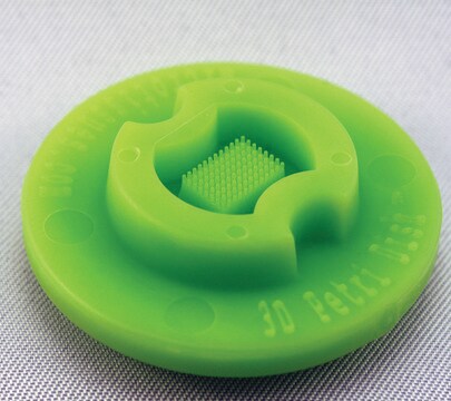
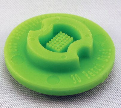
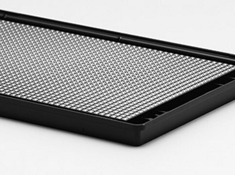
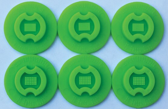
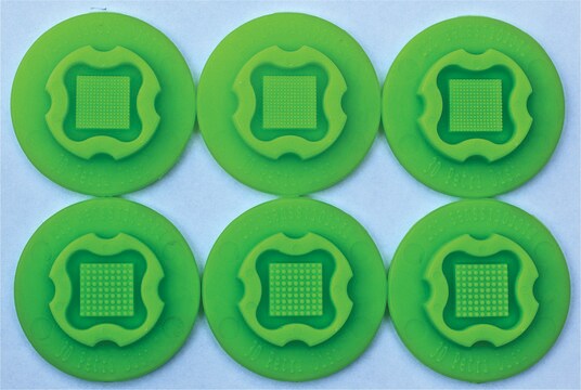
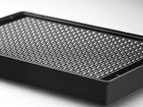
MicroTissues® 3D Petri Dish® micro-mold spheroids
size S, 16 x 16 array, fits 12 well plates
Synonim(y):
3D, 3D Cell Culture
Zaloguj sięWyświetlanie cen organizacyjnych i kontraktowych
About This Item
Kod UNSPSC:
41121812
NACRES:
NB.14
Polecane produkty
Materiały
spherical
rozmiar
S
sterylność
sterile; autoclaved
Właściwości
lid: no
16 x 16 array
opakowanie
pack of 6 ea
producent / nazwa handlowa
MicroTissues Inc. 12-256
pojemność
190 μL
Szukasz podobnych produktów? Odwiedź Przewodnik dotyczący porównywania produktów
Opis ogólny
Six autoclavable precision micro-molds to cast 3D Petri Dish for forming small spheroids. 3D Petri Dish for use in 12-well plate. Each micro-mold forms 256 circular recesses in a 16 x 16 array.
- Nominal dimensions of each 3D culture recess: diam. 300 μm x D 800 μm
- Micro-molds also form single chamber for cell seeding
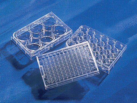
When the gelled agarose is removed from the micro-mold, it is transferred to a standard 12 well or 24 well tissue culture dish and equilibrated with cell culture medium.
Since the agarose is transparent, the spheroids or microtissues that form at the bottom of each agarose micro-well can be easily viewed using a standard inverted microscope using phase contrast, bright field or fluorescence microscopy.
Since the agarose is transparent, the spheroids or microtissues that form at the bottom of each agarose micro-well can be easily viewed using a standard inverted microscope using phase contrast, bright field or fluorescence microscopy.
- The micro-molds are reusable up to 12 times and can be sterilized via a standard steam autoclave (30 min, dry cycle)
- The micro-molds should be stored in a covered container to maintain sterility and to avoid collecting dust or fibres on their small features
- Any microscope with sufficient magnification and resolution functions for cell based views or images will be suitable for use with the 3D Petri Dish products
Informacje prawne
3D Petri Dish is a registered trademark of MicroTissues Inc.
MicroTissues is a registered trademark of MicroTissues Inc.
Ta strona może zawierać tekst przetłumaczony maszynowo.
Wybierz jedną z najnowszych wersji:
Certyfikaty analizy (CoA)
Lot/Batch Number
Przepraszamy, ale COA dla tego produktu nie jest aktualnie dostępny online.
Proszę o kontakt, jeśli potrzebna jest pomoc Obsługa Klienta
Masz już ten produkt?
Dokumenty związane z niedawno zakupionymi produktami zostały zamieszczone w Bibliotece dokumentów.
Klienci oglądali również te produkty
Minjae Kim et al.
Journal of cell science, 132(19) (2019-09-08)
Cultured rat primitive extraembryonic endoderm (pXEN) cells easily form free-floating multicellular vesicles de novo, exemplifying a poorly studied type of morphogenesis. Here, we reveal the underlying mechanism and the identity of the vesicles. We resolve the morphogenesis into vacuolization, vesiculation
Nasz zespół naukowców ma doświadczenie we wszystkich obszarach badań, w tym w naukach przyrodniczych, materiałoznawstwie, syntezie chemicznej, chromatografii, analityce i wielu innych dziedzinach.
Skontaktuj się z zespołem ds. pomocy technicznej
