Przejdź do zawartości
Merck

U3254

Sigma-Aldrich

Monoclonal Anti-Uvomorulin/E-Cadherin antibody produced in rat

clone DECMA-1, ascites fluid, buffered aqueous solution

Synonim(y):

Anti-E-Cadherin, Anti-LCAM

Zaloguj sięWyświetlanie cen organizacyjnych i kontraktowych


About This Item

Numer MDL:
Kod UNSPSC:
12352203
NACRES:
NA.41

pochodzenie biologiczne

rat

Poziom jakości

białko sprzężone

unconjugated

forma przeciwciała

ascites fluid

rodzaj przeciwciała

primary antibodies

klon

DECMA-1, monoclonal

Postać

buffered aqueous solution

zawiera

15 mM sodium azide

reaktywność gatunkowa

bovine, human, canine, mouse

metody

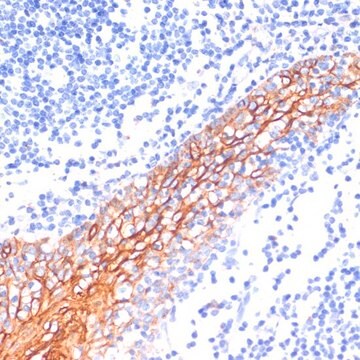
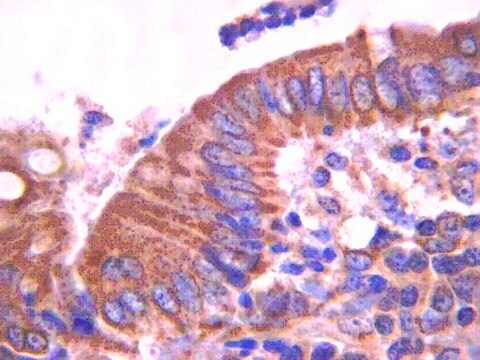
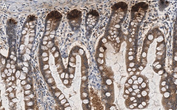
immunohistochemistry (frozen sections): suitable
immunoprecipitation (IP): suitable
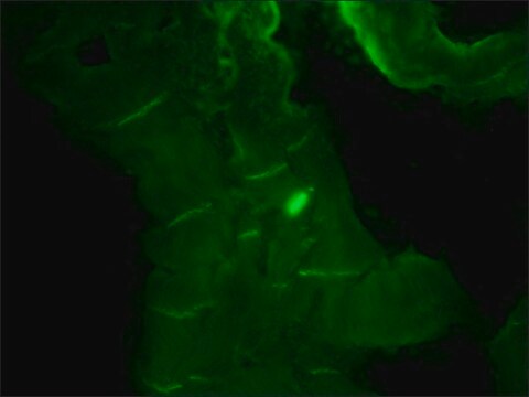
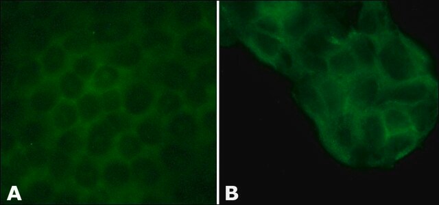
indirect immunofluorescence: 1:1,600 using cultured MDCK cells
microarray: suitable
western blot: suitable

izotyp

IgG1

numer dostępu UniProt

Warunki transportu

dry ice

temp. przechowywania

−20°C

docelowa modyfikacja potranslacyjna

unmodified

informacje o genach

human ... CDH1(999)
mouse ... Cdh1(12550)

Opis ogólny

Monoclonal Anti-Uvomorulin/E-Cadherin (rat IgG1 isotype) is derived from the DECMA-1 hybridoma, produced by the fusion of rat myeloma cells and splenocytes from an immunized Lou rat. Uvomorulin protein was initially identified in embryonal carcinoma and is identical to E-Cadherin, liver-cell adhesion molecules (L-CAM), Cell CAM 80/120, and Activity-regulated cytoskeleton-associated protein 1 (ARC-1), each of which have been characterized in different systems. Uvomorulin/E-Cadherin has been characterized as a 120 kDa cell surface glycoprotein from which an 84 kDa fragment can be released by trypsin digestion in the presence of Ca2+.

Specyficzność

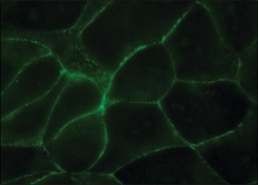
Monoclonal Anti-Uvomorulin/E-Cadherin was selected against the mouse cell adhesion molecule uvomorulin/E-Cadherin. The antibody localizes the cell surface glycoprotein uvomorulin/E-cadherin that has been found to be identical to L-CAM, Cell CAM 80/120, and ARC-1. It blocks both the aggregation of mouse embryonal carcinoma cells and the compaction of pre-implantation embryos. The antibody disrupts confluent monolayers of Madin-Darby canine kidney (MDCK) epithelial cells. In indirect immunofluorescent staining of MDCK cells grown in culture, the antibody shows strong staining on the membrane of adjacent cells, after treatment with 0.5% Triton-X 100.
The antibody localizes the cell surface glycoprotein uvomorulin/E-cadherin that has been found to be identical to L-CAM, Cell CAM 80/120, and ARC-1. The antibody may be used for studies of embryonal development, cell-cell interactions of cultured cells, and localization of uvomorulin/E-cadherin in immunoblotting or immunohistochemical assays.

Immunogen

mouse embryonal carcinoma cell line PCC4 Aza R1.

Zastosowanie

Monoclonal Anti-Uvomorulin/E-Cadherin has been used in immunofluorescence, immunoblotting, immunoprecipitation, immunohistochemistry, macromolecule permeability assay and agglomeration of two embryoid body assay.

Oświadczenie o zrzeczeniu się odpowiedzialności

Unless otherwise stated in our catalog or other company documentation accompanying the product(s), our products are intended for research use only and are not to be used for any other purpose, which includes but is not limited to, unauthorized commercial uses, in vitro diagnostic uses, ex vivo or in vivo therapeutic uses or any type of consumption or application to humans or animals.
This page may contain text that has been machine translated.

Nie możesz znaleźć właściwego produktu?  

Wypróbuj nasz Narzędzie selektora produktów.

Kod klasy składowania

12 - Non Combustible Liquids

Klasa zagrożenia wodnego (WGK)

nwg

Temperatura zapłonu (°F)

Not applicable

Temperatura zapłonu (°C)

Not applicable


Certyfikaty analizy (CoA)

Poszukaj Certyfikaty analizy (CoA), wpisując numer partii/serii produktów. Numery serii i partii można znaleźć na etykiecie produktu po słowach „seria” lub „partia”.

Masz już ten produkt?

Dokumenty związane z niedawno zakupionymi produktami zostały zamieszczone w Bibliotece dokumentów.

Odwiedź Bibliotekę dokumentów

Reduced Pax2 gene dosage increases apoptosis and slows the progression of renal cystic disease
Ostrom L, et al.
Developmental Biology, 219(2), 250-258 (2000)
Controlled, scalable embryonic stem cell differentiation culture
Dang SM, et al.
Stem Cells, 22(3), 275-282 (2004)
Claudin-3 overexpression increases the malignant potential of colorectal cancer cells: roles of ERK1/2 and PI3K-Akt as modulators of EGFR signaling
de Souza WF, et al.
PLoS ONE, 8(9), e74994-e74994 (2013)
TGF-beta induced transdifferentiation of mammary epithelial cells to mesenchymal cells: involvement of type I receptors.
Miettinen PJ, et al.
The Journal of Cell Biology, 127(6), 2021-2036 (1994)
Karen Rodgers et al.
The American journal of pathology, 167(3), 683-694 (2005-08-30)
Lipoxins (LXs), endogenously produced eicosanoids, possess potent anti-inflammatory, pro-resolution bioactivities. We investigated the potential of LXA(4) (1 to 10 nmol/L) to modify the effects of platelet-derived growth factor (PDGF)-induced gene expression in human renal mesangial cells (hMCs). Using oligonucleotide microarray

Nasz zespół naukowców ma doświadczenie we wszystkich obszarach badań, w tym w naukach przyrodniczych, materiałoznawstwie, syntezie chemicznej, chromatografii, analityce i wielu innych dziedzinach.

Skontaktuj się z zespołem ds. pomocy technicznej