
PICM01250
Millicell® Standing Cell Culture Inserts
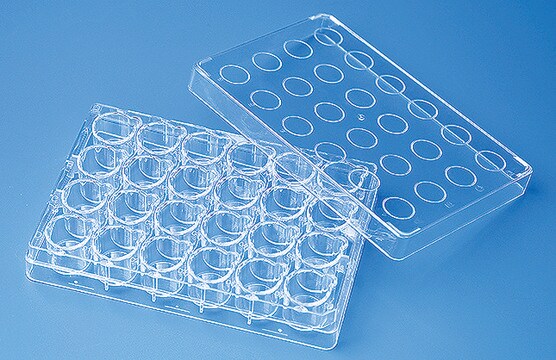
pore size 0.4 μm, diam. 12 mm, transparent PTFE membrane, hydrophilic, size 24 wells, sterile
Synonim(y):
Millicell Cell Culture Insert, 12 mm, hydrophilic PTFE, 0.4 µm, Millicell-CM, cell culture inserts, permeable culture insert, plate inserts, tissue culture insert, tissue culture plate
About This Item
Polecane produkty

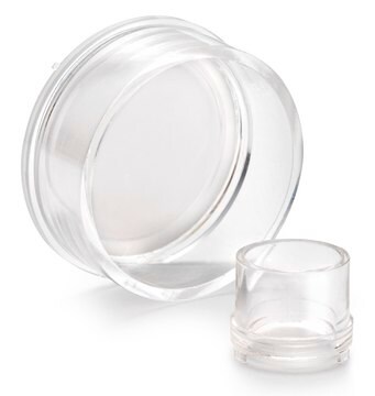


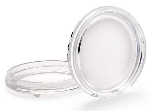
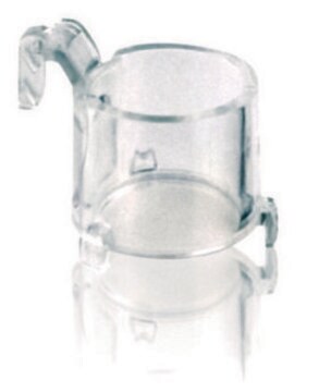
Materiały
polystyrene housing
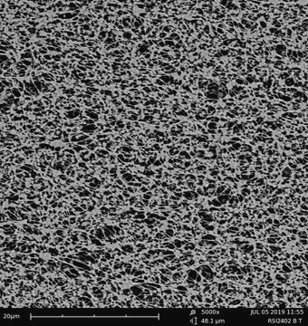
transparent PTFE membrane
Poziom jakości
sterylność
ethylene oxide treated
sterile
Właściwości
hydrophilic
opakowanie
pack of 50 (individually blister packaged)
producent / nazwa handlowa
Millicell®
Parametry
50 °C max. temp.
metody
cell attachment: suitable
cell culture | mammalian: suitable
cell differentiation: suitable
Wysokość
10.5 mm
Średnica
12 mm
powierzchnia filtracyjna
0.6 cm2
rozmiar
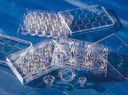
24 wells
powierzchnia
0.6 cm2
pojemność robocza
0.6 mL
kolor
transparent membrane, when wetted
Matryca
Biopore™
wielkość porów
0.4 μm
typ wiązania
low binding surface
metoda wykrywania
fluorometric
Warunki transportu
ambient
Opis ogólny
Zastosowanie
- Cell Attachment
- Cell Growth
- Cell Differentiation
- Immunocytochemistry
Cechy i korzyści
- Promotes excellent cell growth and provides an exceptional opportunity for cell studies
- Low protein-binding hydrophilic PTFE membrane is ideal for live cell viewing and immunofluorescent application
Informacje prawne
polecane
produkt powiązany
wyposażenie dodatkowe
Kod klasy składowania
10-13 - German Storage Class 10 to 13
Certyfikaty analizy (CoA)
Poszukaj Certyfikaty analizy (CoA), wpisując numer partii/serii produktów. Numery serii i partii można znaleźć na etykiecie produktu po słowach „seria” lub „partia”.
Masz już ten produkt?
Dokumenty związane z niedawno zakupionymi produktami zostały zamieszczone w Bibliotece dokumentów.
Klienci oglądali również te produkty

Produkty
16HBE14o- ludzkie komórki nabłonka oskrzeli wykorzystywane do modelowania nabłonka oddechowego w badaniach nad mukowiscydozą, wirusową patologią płuc (SARS-CoV), astmą, POChP, skutkami palenia tytoniu i zanieczyszczenia powietrza. Zobacz ponad 5 tys. publikacji.
Protokoły
This is a Toluidine Blue Staining protocol.
Błękit toluidynowy selektywnie barwi materiał jądrowy i kwaśne składniki tkanek, pomagając w barwieniu histologicznym tkanek bogatych w DNA/RNA.
3D cell culture protocol for generating epidermal human skin tissue using primary human keratinocytes, dermal fibroblasts, and collagen-coated transwell inserts.
This protocol covers 3 modes for the microscopic examination of cell samples.
Powiązane treści
This page covers the ECM coating protocols developed for four types of ECMs on Millicell®-CM inserts, Collagen Type 1, Fibronectin, Laminin, and Matrigel.
Nasz zespół naukowców ma doświadczenie we wszystkich obszarach badań, w tym w naukach przyrodniczych, materiałoznawstwie, syntezie chemicznej, chromatografii, analityce i wielu innych dziedzinach.
Skontaktuj się z zespołem ds. pomocy technicznej