Przejdź do zawartości
Merck
Wszystkie zdjęcia(2)

Kluczowe dokumenty

MAB8151-I

Sigma-Aldrich

Anti-West Nile Virus/Kunjin Antibody, Envelope Antibody, clone 3.91D

Zaloguj sięWyświetlanie cen organizacyjnych i kontraktowych


About This Item

Kod UNSPSC:
12352203
NACRES:
NA.41

pochodzenie biologiczne

mouse

Poziom jakości

białko sprzężone

unconjugated

forma przeciwciała

purified antibody

rodzaj przeciwciała

primary antibodies

klon

3.91D, monoclonal

masa cząsteczkowa

calculated mol wt 31.89 kDa
observed mol wt ~50 kDa

oczyszczone przez

using protein G

reaktywność gatunkowa

virus

opakowanie

antibody small pack of 100 μL

metody

ELISA: suitable
immunofluorescence: suitable
western blot: suitable

izotyp

IgG3

sekwencja epitopowa

N-terminal half

numer dostępu Protein ID

numer dostępu UniProt

Warunki transportu

2-8°C

docelowa modyfikacja potranslacyjna

unmodified

informacje o genach

vaccinia virus ... poly> POLY(912267)

Opis ogólny

Genome polyprotein (UniProt: P14335) is encoded by the POLY gene (Gene ID: 912267) in West Nile virus. West Nile virus (WNV) is a mosquito-borne positive-sense RNA virus of the Flavivirus genus within the Flaviviridae family. WNV genome encodes for three structural domains known as C, prM, and E and seven non-structural proteins (NS1, NS2A, NS2B, NS3, NS4A, NS4B, and NS5). These are translated as a single polypeptide, which is subsequently cleaved by viral and cellular proteases. The envelope glycoprotein (E) undergoes a conformational rearrangement triggered by low pH that results in a class II fusion event required for viral entry. The E protein (aa 291-791) contains a three-domain structure where domain I is an eight-stranded β-barrel in the center portion of the protein (aa 1-51, 134-195, and 284-297). The DII domain consists of 170 residues inserted into DI (aa 52-133 and 196-283). At the distal end of DII domain is a fusion loop (aa 98-110) and a conserved glycine-rich, hydrophobic sequence, which is important for flaviviral fusion. The third domain, DIII, is an Ig-like domain with seven β-strand and has been implicated in receptor binding. The E protein also has a single N-linked glycosylation site at Asn 154, located within the ectodomain of prM domain and is cleaved by cellular enzymes during viral particle maturation. Clone E60 specifically binds to the DII fusion loop and displays limited neutralizing activity and even at higher doses neutralization activity is shown to be between 26 and 68%. (Ref.: Martín-Acebes MA, et al. (2012). World J Virol. 1(2); 51-70).

Specyficzność

Clone 3.91D is a mouse monoclonal antibody that detects West Nile virus envelope protein. It targets an epitope within the N-terminal half of polyprotein.

Immunogen

West Nile Virus (WNV; subtype KUNV) isolate OR393.

Zastosowanie

Qualtiy Control Testing

Evaluated by Western Blotting with recombinant West Nile Virus envelope protein.

Western Blotting Analysis (WB): A 1:500 dilution of this antibody detected recombinant West Nile Virus envelope protein.

Tested Applications


Western Blotting Analysis: A representative lot detected West Nile Virus/Kunjin, Envelope protein in Western Blotting applications (Maeda, A., et al. (2009). Virus Res.;144(1-2):35-43; Saiyasombat, R., et al. (2014). Virol J.;11:150; Blitvich, B.J., et al. (2016). Am J Trop Med Hyg.;95(5):1185-1191).

ELISA Analysis: Various dilutions of this antibody detected recombinant West Nile Virus/Kunjin, Envelope protein.

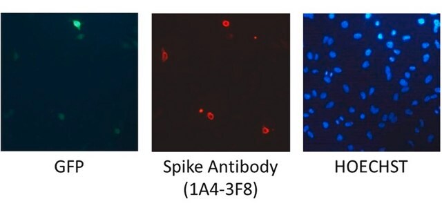
Immunofluorescence Analysis: A representative lot detected West Nile Virus/Kunjin, Envelope protein in Immunofluorescence applications (Maeda, A., et al. (2009). Virus Res.;144(1-2):35-43; Osorio, J.E., et al. (2012). Am J Trop Med Hyg.;87(3):565-72).

Note: Actual optimal working dilutions must be determined by end user as specimens, and experimental conditions may vary with the end user
Anti-West Nile Virus/Kunjin, Envelope, clone 3.91D, Cat. No. MAB8151-I, is a mouse monoclonal antibody that detects envelope protein in West Nile virus and is tested for use in ELISA, Immunofluorescence, and Western Blotting.

Postać fizyczna

Purified mouse monoclonal antibody IgG3 in buffer containing 0.1 M Tris-Glycine (pH 7.4), 150 mM NaCl with 0.05% sodium azide.

Przechowywanie i stabilność

Recommend storage at +2°C to +8°C. For long term storage antibodies can be kept at -20°C. Avoid repeated freeze-thaws.

Inne uwagi

Concentration: Please refer to the Certificate of Analysis for the lot-specific concentration.

Oświadczenie o zrzeczeniu się odpowiedzialności

Unless otherwise stated in our catalog or other company documentation accompanying the product(s), our products are intended for research use only and are not to be used for any other purpose, which includes but is not limited to, unauthorized commercial uses, in vitro diagnostic uses, ex vivo or in vivo therapeutic uses or any type of consumption or application to humans or animals.
Ta strona może zawierać tekst przetłumaczony maszynowo.

Nie możesz znaleźć właściwego produktu?  

Wypróbuj nasz Narzędzie selektora produktów.

Kod klasy składowania

12 - Non Combustible Liquids

Klasa zagrożenia wodnego (WGK)

WGK 1

Temperatura zapłonu (°F)

Not applicable

Temperatura zapłonu (°C)

Not applicable


Certyfikaty analizy (CoA)

Poszukaj Certyfikaty analizy (CoA), wpisując numer partii/serii produktów. Numery serii i partii można znaleźć na etykiecie produktu po słowach „seria” lub „partia”.

Masz już ten produkt?

Dokumenty związane z niedawno zakupionymi produktami zostały zamieszczone w Bibliotece dokumentów.

Odwiedź Bibliotekę dokumentów

Nasz zespół naukowców ma doświadczenie we wszystkich obszarach badań, w tym w naukach przyrodniczych, materiałoznawstwie, syntezie chemicznej, chromatografii, analityce i wielu innych dziedzinach.

Skontaktuj się z zespołem ds. pomocy technicznej