コンテンツへスキップ
Merck

PKH26PCL

Sigma-Aldrich

PKH26赤色蛍光細胞リンカーキット, 食細胞標識用

Distributed for Phanos Technologies

別名:

Phagocytic cell label

ログイン組織・契約価格を表示する


About This Item

UNSPSCコード:
12352207
NACRES:
NA.32

アプリケーション

本キットは貪食細胞の標識用です。単球、マクロファ-ジ、好中球のような食作用を持つ細胞を選択的に標識するのに用います。

原理

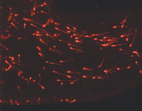
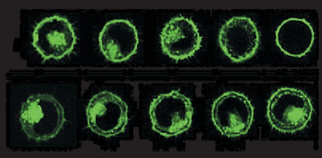
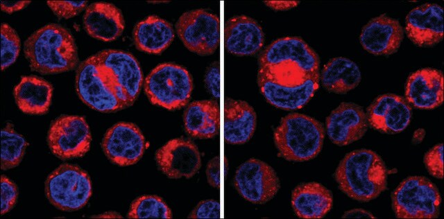
色素が凝集、微粒子を形成して標識がおこります。色素凝集体が形成されるとリンパ球など非食細胞による色素取り込みは大きく阻害されますが、食細胞による色素取り込みが促進します。色素は細胞内の貪食部分に局在するため、標識細胞は斑点状またはスポット状に観察されます。色素は代謝されにくく、in vivoにおいて細胞内に21日以上留まることが確認されています。
in vitroin vivoのどちらでも貪食細胞の標識が可能です。in vivo ではPKH26標識溶液を腹腔内または静脈内に注射し食細胞を標識します。分離した標的細胞の場合はin vitro標識法で染色できます。

関連事項

他の多くの論文を含めた、PKH および CellVue® 蛍光細胞リンカー色素に関する追加の技術詳細については、こちらをご覧ください。

法的情報

CellVue is a registered trademark of Phanos Technologies

キットの構成要素のみ

製品番号
詳細

  • Diluent B 6 x 10

  • PKH26 cell linker in ethanol .5 mL

関連製品

ピクトグラム

FlameExclamation mark

シグナルワード

Danger

危険有害性情報

危険有害性の分類

Eye Irrit. 2 - Flam. Liq. 2

保管分類コード

3 - Flammable liquids

引火点(°F)

57.2 °F - closed cup

引火点(℃)

14.0 °C - closed cup


適用法令

試験研究用途を考慮した関連法令を主に挙げております。化学物質以外については、一部の情報のみ提供しています。 製品を安全かつ合法的に使用することは、使用者の義務です。最新情報により修正される場合があります。WEBの反映には時間を要することがあるため、適宜SDSをご参照ください。

毒物及び劇物取締法

キットコンポーネントの情報を参照してください

PRTR

キットコンポーネントの情報を参照してください

消防法

キットコンポーネントの情報を参照してください

労働安全衛生法名称等を表示すべき危険物及び有害物

キットコンポーネントの情報を参照してください

労働安全衛生法名称等を通知すべき危険物及び有害物

キットコンポーネントの情報を参照してください

カルタヘナ法

キットコンポーネントの情報を参照してください

Jan Code

キットコンポーネントの情報を参照してください


試験成績書(COA)

製品のロット番号・バッチ番号を入力して、試験成績書(COA) を検索できます。ロット番号・バッチ番号は、製品ラベルに「Lot」または「Batch」に続いて記載されています。

以前この製品を購入いただいたことがある場合

文書ライブラリで、最近購入した製品の文書を検索できます。

文書ライブラリにアクセスする

Hyung-Syup Kim et al.
Journal of Korean Neurosurgical Society, 44(4), 249-255 (2008-12-20)
In Moyamoya disease, the primary goal of treatment is to improve collateral circulation through angiogenesis. In the present study, we obtained and sub-cultured bone marrow stromal cells (BMSCs) from rats without a cell-mediated immune response. Then, we injected the labeled
Jaime Murphy et al.
American journal of respiratory cell and molecular biology, 38(4), 380-385 (2008-01-15)
To further examine the half-life of alveolar macrophages, chimeric CD 45.2 mice were generated through bone marrow transplantation of donor CD 45.1 cells. Before administration of donor cells, recipient mice were divided into two cohorts: the first cohort received total
K Tabata et al.
Gene therapy, 18(10), 969-978 (2011-04-23)
We previously identified the mouse and human Glipr1 and GLIPR1/RTVP-1 genes, respectively, as direct p53 targets with proapoptotic activities in various cancer cell lines, including prostate cancer (PCa). Intratumoral injection of an adenoviral vector capable of efficient transduction and expression
Lara Campana et al.
Journal of immunology (Baltimore, Md. : 1950), 200(3), 1169-1187 (2017-12-22)
The disposal of apoptotic bodies by professional phagocytes is crucial to effective inflammation resolution. Our ability to improve the disposal of apoptotic bodies by professional phagocytes is impaired by a limited understanding of the molecular mechanisms that regulate the engulfment
Meaghan M Hunter et al.
Gastroenterology, 138(4), 1395-1405 (2010-01-05)
Infection with the rat tapeworm Hymenolepis diminuta reduces the severity of dinitrobenzene sulfonic acid (DNBS)-induced colitis in mice. Infection with H. diminuta increases colonic expression of arginase-1 and found in inflammatory zone 1 (FIZZ1), markers of alternatively activated macrophages (AAMs).

資料

PKH dyes are easy to use and achieve stable, uniform, and reproducible fluorescent labeling of live cells. PKH dyes are non-toxic membrane stains which produce high signal to noise ratio.

Lipophilic cell tracking dyes enable cancer biologists to track tumor and immune cell functions both in vitro and in vivo. Read the article to choose a right membrane dye kit for cell tracking and proliferation monitoring.

Optimal staining is a key component for studying tumorigenesis and progression. Learn useful tips and techniques for dye applications, including examples from recent studies.

A video about how you can use fluorescent cell tracking dyes in combination with flow and image cytometry to study interactions and fates of different cell types in vitro and in vivo.

すべて表示

ライフサイエンス、有機合成、材料科学、クロマトグラフィー、分析など、あらゆる分野の研究に経験のあるメンバーがおります。.

製品に関するお問い合わせはこちら(テクニカルサービス)