おすすめの製品
品質水準
包装
pkg of 1 kit
保管条件
protect from light
蛍光検出
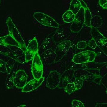
λex 490 nm; λem 502 nm (PKH67 dye)
検出方法
fluorometric
輸送温度
ambient
保管温度
room temp
アプリケーション
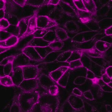
- トリプルネガティブ乳がん病態の細胞から放出されたエキソソームを標識し、さらに調べることに。
- アポトーシス細胞を標識するために。アポトーシス細胞の結合および内在化の両方における ανβ5受容体の役割を研究するために。
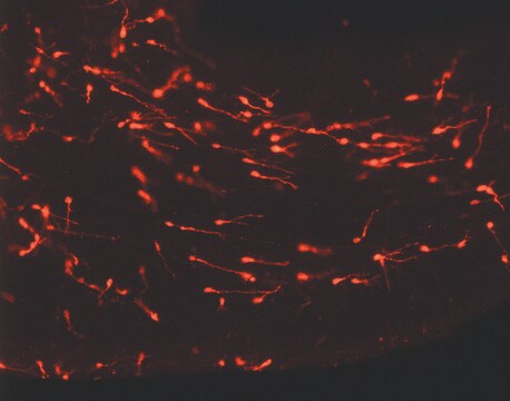
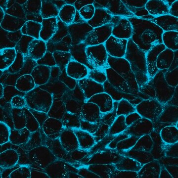
- 蛍光画像法で。
PKH1およびPKH2を用いたin vivo 研究では、蛍光の消失が遅いことが観察されています。 この挙動は緑色細胞リンカ-色素に特徴的ですが、赤色細胞リンカ-色素には特徴的でないことから、PKH67も同様の性質を示すと考えられます。非分裂細胞におけるin vitro細胞膜維持率とin vivo蛍光半減期との相関から、PKH67のin vivo蛍光半減期は10~12日と予測されます。同程度の半減期を持つ他の緑色蛍光リンカ-色素は、1-2カ月間のin vivo リンパ球およびマクロファ-ジ輸送の観察に使用されていることから、PKH67も適度な期間のin vivo トラッキング研究に使用できると考えられます。
関連事項
法的情報
キットの構成要素のみ
- Diluent C 6 x 10
- PKH67 Cell Linker in ethanol .5 mL
シグナルワード
Danger
危険有害性情報
危険有害性の分類
Eye Irrit. 2 - Flam. Liq. 2
保管分類コード
3 - Flammable liquids
引火点(°F)
57.2 °F - closed cup
引火点(℃)
14 °C - closed cup
適用法令
試験研究用途を考慮した関連法令を主に挙げております。化学物質以外については、一部の情報のみ提供しています。 製品を安全かつ合法的に使用することは、使用者の義務です。最新情報により修正される場合があります。WEBの反映には時間を要することがあるため、適宜SDSをご参照ください。
毒物及び劇物取締法
キットコンポーネントの情報を参照してください
PRTR
キットコンポーネントの情報を参照してください
消防法
キットコンポーネントの情報を参照してください
労働安全衛生法名称等を表示すべき危険物及び有害物
キットコンポーネントの情報を参照してください
労働安全衛生法名称等を通知すべき危険物及び有害物
キットコンポーネントの情報を参照してください
カルタヘナ法
キットコンポーネントの情報を参照してください
Jan Code
キットコンポーネントの情報を参照してください
試験成績書(COA)
製品のロット番号・バッチ番号を入力して、試験成績書(COA) を検索できます。ロット番号・バッチ番号は、製品ラベルに「Lot」または「Batch」に続いて記載されています。
この製品を見ている人はこちらもチェック
資料
PKH dyes are easy to use and achieve stable, uniform, and reproducible fluorescent labeling of live cells. PKH dyes are non-toxic membrane stains which produce high signal to noise ratio.
Lipophilic cell tracking dyes enable cancer biologists to track tumor and immune cell functions both in vitro and in vivo. Read the article to choose a right membrane dye kit for cell tracking and proliferation monitoring.
Optimal staining is a key component for studying tumorigenesis and progression. Learn useful tips and techniques for dye applications, including examples from recent studies.
A video about how you can use fluorescent cell tracking dyes in combination with flow and image cytometry to study interactions and fates of different cell types in vitro and in vivo.
ライフサイエンス、有機合成、材料科学、クロマトグラフィー、分析など、あらゆる分野の研究に経験のあるメンバーがおります。.
製品に関するお問い合わせはこちら(テクニカルサービス)