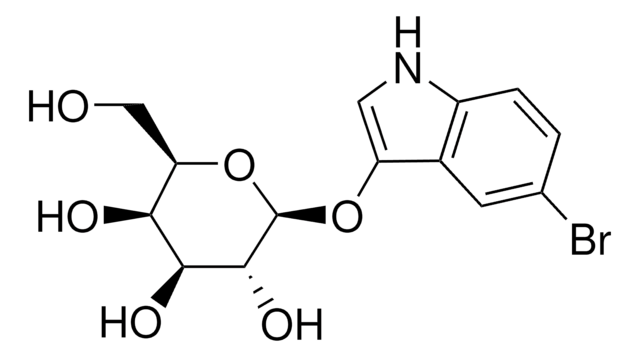
94433
β-Galactosidase stain
別名:
6′-(Diethylamino)-4′-(fluoromethyl)spiro[isobenzofuran-1(3H),9′-[9H]xanthen]-3′-yl β-D-galactopyranoside, SPiDER-βGal, SPiDER-beta Gal, (2S,3R,4S,5R,6R)-2-{[3’-(Diethylamino)-5’-(fluoromethyl)-3H-spiro(isobenzofuran-1,9’-xanthen)-6’-yl]oxy}-6-(hydroxymethyl)tetrahydro-2H-pyran-3,4,5-triol
ログイン組織・契約価格を表示する
すべての画像(1)
About This Item
おすすめの製品
フォーム
solid
品質水準
保管温度
2-8°C
SMILES記法
O[C@@H]1[C@@H](O)[C@H](OC(C=C2)=C(CF)C3=C2C4(C(C=CC=C5)=C5CO4)C(C=CC(N(CC)CC)=C6)=C6O3)O[C@H](CO)[C@@H]1O
InChI
1S/C31H34FNO8/c1-3-33(4-2)18-9-10-21-24(13-18)39-29-19(14-32)23(40-30-28(37)27(36)26(35)25(15-34)41-30)12-11-22(29)31(21)20-8-6-5-7-17(20)16-38-31/h5-13,25-28,30,34-37H,3-4,14-16H2,1-2H3/t25-,26+,27+,28-,30-,31?/m1/s1
InChI Key
SVEDIEUQTRCORD-JGANWENXSA-N
詳細
The gene of β-galactosidase from E. coli is widely used as a reporter gene assay marker. Although X-gal is well known reagent to detect β-galactosidase in cell or tissue samples, the assay using these reagents require to fix cells or tissues due to the poor cell-permeability. In addition, so far developed the assay using fluorescence reagents can not clearly differentiate β-galactosidase-expressed cells or regions.
To overcome these issues, Urano, Kamiya and co-workers have successfully developed a stain ideally possesses cell-permeability and the ability to retain in intracellular region.1)
By the enzymatic reaction, the β-Galactosidase stain immediately forms a quinone methide that acts as electrophile when proteins containing nucleophilic functional groups nearby the molecules. By the probe undergoes the reaction with a protein, the conjugates become fluorescent compounds. Thus, it allows a single-cell analysis because it does self-immobilizing to the intracellular proteins.
Exciation Maximum: 530 nm (± 10 nm)
Emission Maximum: 550 nm (± 10 nm)
Reference:
1) Y. Urano, M. Kamiya, T. Doura, WO 2015174460, A1, (19, November, 2015).
Find more infomation here 94433
アプリケーション
Preparation of 1 mmol stain DMSO stock solution
Add 35 μl of DMSO to a tube of stain solution (20 μg) and dissolve it with pipetting.
*Store the stock solution at -20°C.
Preparation of 1 μmol/l stain working solution
Dilute the stain DMSO stock solution with Hanks′ HEPES buffer to prepare 1 μmol/l stain working solution.
*Hanks′ HEPES buffer is recommended to maintain cell condition.
General protocol:
staining
1. Prepare cells for the assay.
2. Discard the culture medium and wash the cells with Hanks′ HEPES buffer twice.
3. Add an appropriate volume of stain working solution.
4. Incubate at 37oC for 15 minutes.
5. Observe the cells under a fluorescence microscope or by a flow cytometer.
*After staining, the cells can be observed even without washing. However, you can wash it as needed.
保管分類コード
11 - Combustible Solids
WGK
WGK 3
この製品を見ている人はこちらもチェック
Dongqin Yang et al.
EBioMedicine, 47, 114-127 (2019-09-10)
There are many reports of the anti-tumour effects of exogenous adenosine in gastrointestinal tumours. Gemcitabine, a first line agent for patients with poor performance status, and adenosine have structural similarities. For these reasons, it is worth exploring the therapeutic efficacy
ライフサイエンス、有機合成、材料科学、クロマトグラフィー、分析など、あらゆる分野の研究に経験のあるメンバーがおります。.
製品に関するお問い合わせはこちら(テクニカルサービス)
