NS220
Neurite Outgrowth Assay Kit (3 µm)
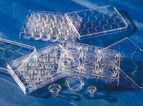
The NS220 Neurite Outgrowth Assay Kit (3 µm) is based on the use of Millicell cell culture inserts (chambers) containing a permeable membrane with 3-μm pores at the base.
Sinônimo(s):
3 µm Assay Kit, Assay Kit for Neurites, Neurite Outgrowth Kit
About This Item
Produtos recomendados
Nível de qualidade
reatividade da espécie (prevista por homologia)
all
fabricante/nome comercial
Chemicon®
técnica(s)
cell based assay: suitable
método de detecção
colorimetric
Condições de expedição
wet ice
Descrição geral
CHEMICON′s NS220 Neurite Outgrowth Assay Kit (3 µm) utilizes microporous tissue culture insert technology from Millipore and is based on the use of Millicell cell culture inserts (chambers) containing a permeable membrane with 3-μm pores at the base. The Millicell inserts utilized in this kit are appropriate for cells projecting neurites of up to 3 µm diameter, e.g. N1E-115 cells [Abe et al., 2003], Dorsal Root Ganglia and Schwann cells [Windebank et al., 1985]. The inserts are designed to fit into a receiver vessel, which when in use contains differentiation media in contact with the bottom of the insert membrane. The permeable membranes allow for discrimination between neurites and cell bodies, as projecting neurites will pass easily through the pores but cell bodies will not. Therefore, by inducing neurites to traverse these membrane pores, a purified population of neurites is located on the underside of the membrane, distal to the surface on which neural cell bodies are deposited.
The assay is a simple, efficient, and versatile system for the quantitative determination of compounds that influence neurite formation and repulsion. With this system, it is possible to screen numerous biological and pharmacological agents simultaneously, directly evaluate adhesion and guidance receptor functions responsible for neurite extension and repulsion, as well as analyze gene function in transfected cells. The microporous filter allows for biochemical separation and purification of neurites and cell bodies for detailed molecular analysis of protein expression, signal transduction processes and identification of drug targets that regulate neurite outgrowth or retraction processes. Reagents and materials supplied in the NS220 Neurite Outgrowth Assay Kit are sufficient for 12 tests.
Note: the 3 µm pore size inserts in this kit are unsuitable for use with PC12 cells. Due to the relatively small diameter of PC12 cell bodies [Abe et al., 2003], some of these cells may pass though 3 µm pores and obscure the results. Chemicon′s NS225 Neurite Outgrowth Assay Kit (1 µm) has been specifically designed for and validated with PC12 cells.
Aplicação
Componentes
2. Neurite Stain Solution: (Part No. 90242) One 20 mL bottle.
3. Neurite Stain Extraction Buffer: (Part No. 90243) One 20 mL bottle.
4. Neurite Outgrowth Assay Plate: (Part No. 2007255) Two 24-well plates.
5. Cotton Swabs: (Part No. 10202) 50 swabs.
6. Forceps: (Part No. 10203) One each.
Armazenamento e estabilidade
Informações legais
Exoneração de responsabilidade
Palavra indicadora
Danger
Frases de perigo
Declarações de precaução
Classificações de perigo
Eye Irrit. 2 - Flam. Liq. 2
Código de classe de armazenamento
3 - Flammable liquids
Certificados de análise (COA)
Busque Certificados de análise (COA) digitando o Número do Lote do produto. Os números de lote e remessa podem ser encontrados no rótulo de um produto após a palavra “Lot” ou “Batch”.
Já possui este produto?
Encontre a documentação dos produtos que você adquiriu recentemente na biblioteca de documentos.
Artigos
Optimized cell based neurite outgrowth assays and reagents to study neuron function and development.
Nossa equipe de cientistas tem experiência em todas as áreas de pesquisa, incluindo Life Sciences, ciência de materiais, síntese química, cromatografia, química analítica e muitas outras.
Entre em contato com a assistência técnica


