MABF39-I
Anti-DC-STAMP Antibody, clone 1A2
clone 1A2, from mouse
Sinônimo(s):
Dendritic cell-specific transmembrane protein, DC-STAMP, Dendrocyte-expressed seven transmembrane protein, FIND, hDC-STAMP, IL-four-induced protein, Transmembrane 7 superfamily member 4
About This Item
Produtos recomendados
fonte biológica
mouse
Nível de qualidade
forma do anticorpo
purified immunoglobulin
tipo de produto de anticorpo
primary antibodies
clone
1A2, monoclonal
reatividade de espécies
mouse, human
técnica(s)
flow cytometry: suitable
immunocytochemistry: suitable
immunoprecipitation (IP): suitable
western blot: suitable
Isotipo
IgG2aκ
nº de adesão NCBI
nº de adesão UniProt
modificação pós-traducional do alvo
unmodified
Informações sobre genes
human ... DCSTAMP (81501)
Descrição geral
Especificidade
Imunogênio
Aplicação
Inflammation & Immunology
Osteobiology
Western Blotting Analysis: 1.0 µg/mL from a representative lot detected DC-STAMP in thymus and spleen lysates from wild-type mice, and greatly reduced DC-STAMP in thymus and spleen lysates from Dcstamp-knockout mice (Courtesy of Grace Chiu, Ph.D., University of Rochester Medical Center, NY, USA).
Western Blotting Analysis: A representative lot detected the ~106 kDa dimeric and the ~53 kDa monomeric DC-STAMP band by Western blotting under non-denatured and denatured condition, respectively, following DC-STAMP immunoprecipitation using murine RAW 264.7 macrophage lysate (Mensah, K.A., et al. (2010). J. Cell. Physiol. 223(1):76-83).
Flow Cytometry Analysis: A representative lot detected DC-STAMP-positive lymphoctes and monocytes in purified human PBMCs (Chiu, Y.H., et al. (2012). J. Bone Miner. Res. 27(1):79-92).
Flow Cytometry Analysis: A representative lot detected DC-STAMP surface expression on murine RAW 264.7 macrophages and murine bone marrow-derived CD11b+ monocytes. DC-STAMP is expressed on osteoclast precursor (OCP) cells as a dimer, which is efficiently detected by flow cytometry using clone 1A2 (Mensah, K.A., et al. (2010). J. Cell. Physiol. 223(1):76-83).
Function Analysis: A representative lot inhibited RANKL & M-CSF treatment-induced osteoclasts (OC) formation in human PBMCs & monocytes cultures (Chiu, Y.H., et al. (2012). J. Bone Miner. Res. 27(1):79-92).
Function Analysis: A representative lot inhibited RANKL treatment-induced osteoclasts (OC) formation in RAW 264.7 and murine bone marrow macrophages cultures (Mensah, K.A., et al. (2010). J. Cell. Physiol. 223(1):76-83).
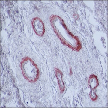
Immunohistochemistry Analysis: A representative lot detected DC-STAMP expressionon in multinucleated ‘osteoclast-like’ giant cells in human giant cell tumor of bone (Chiu, Y.H., et al. (2012). J. Bone Miner. Res. 27(1):79-92).
Immunocytochemistry Analysis: A representative lot detected DC-STAMP-positive human PBMCs using 10% NBF-fixed, paraffin-embedded cell preparation (Chiu, Y.H., et al. (2012). J. Bone Miner. Res. 27(1):79-92).
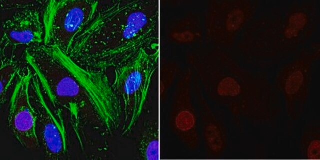
Immunocytochemistry Analysis: A representative lot detected differential DC-STAMP intracellular localizations by fluorescent immunocytochemistry staining of 4% paraformaldehyde-fixed, 0.1% saponin-permeabilized murine bone marrow macrophages at different time points during osteoclastogenesis upon RANKL treatment (Mensah, K.A., et al. (2010). J. Cell. Physiol. 223(1):76-83).
Immunoprecipitation Analysis: A representative lot immunoprecipitated DC-STAMP from the lysates of human monocytes (Chiu, Y.H., et al. (2012). J. Bone Miner. Res. 27(1):79-92).
Immunoprecipitation Analysis: A representative lot immunoprecipitated DC-STAMP from the membrane extracts of murine RAW 264.7 macrophages (Mensah, K.A., et al. (2010). J. Cell. Physiol. 223(1):76-83).
Qualidade
Western Blotting Analysis: 4.0 µg/mL of this antibody detected DC-STAMP in 10 µg of mouse thymus lysate.
Descrição-alvo
forma física
Armazenamento e estabilidade
Handling Recommendations: Upon receipt and prior to removing the cap, centrifuge the vial and gently mix the solution. Aliquot into microcentrifuge tubes and store at -20°C. Avoid repeated freeze/thaw cycles, which may damage IgG and affect product performance.
Outras notas
Exoneração de responsabilidade
Não está encontrando o produto certo?
Experimente o nosso Ferramenta de seleção de produtos.
Código de classe de armazenamento
12 - Non Combustible Liquids
Classe de risco de água (WGK)
WGK 1
Ponto de fulgor (°F)
Not applicable
Ponto de fulgor (°C)
Not applicable
Certificados de análise (COA)
Busque Certificados de análise (COA) digitando o Número do Lote do produto. Os números de lote e remessa podem ser encontrados no rótulo de um produto após a palavra “Lot” ou “Batch”.
Já possui este produto?
Encontre a documentação dos produtos que você adquiriu recentemente na biblioteca de documentos.
Nossa equipe de cientistas tem experiência em todas as áreas de pesquisa, incluindo Life Sciences, ciência de materiais, síntese química, cromatografia, química analítica e muitas outras.
Entre em contato com a assistência técnica


