Pular para o conteúdo
Merck
Todas as fotos(1)

Documentos Principais

MAB351R

Sigma-Aldrich

Anti-GAD2(GAD67) Antibody

CHEMICON®, mouse monoclonal, GAD-6

Sinônimo(s):

GAD65

Faça loginpara ver os preços organizacionais e de contrato


About This Item

Código UNSPSC:
12352203
eCl@ss:
32160702
NACRES:
NA.41

Nome do produto

Anti-Glutamate Decarboxylase Antibody, 65 kDa isoform, clone GAD-6, clone GAD-6, Chemicon®, from mouse

fonte biológica

mouse

Nível de qualidade

forma do anticorpo

purified immunoglobulin

tipo de produto de anticorpo

primary antibodies

clone

GAD-6, monoclonal

reatividade de espécies

human, rat

fabricante/nome comercial

Chemicon®

técnica(s)

immunohistochemistry: suitable
western blot: suitable

Isotipo

IgG2a

nº de adesão NCBI

nº de adesão UniProt

Condições de expedição

dry ice

modificação pós-traducional do alvo

unmodified

Informações sobre genes

human ... GAD2(2572)

Descrição geral

Gutamic acid decarboxylase (GAD; E.C. 4.1.1.15) is the enzyme responsible for the conversion of glutamic acid to gamma-aminobutyric acid (GABA), the major inhibitory transmitter in higher brain regions, and putative paracrine hormone in pancreatic islets. Two molecular forms of GAD (65kDa and 67kDa, 64% aa identity between forms) are highly conserved and both forms are expressed in the CNS, pancreatic islet cells, testis, oviduct and ovary. The isoforms are regionally distributed cytoplasmically in the brains of rats and mice (Sheikh, S. et al. 1999). GAD65 is an ampiphilic, membrane-anchored protein (585aa), encoded on human chromosome 10, and is responsible for vesicular GABA production. GAD67 is cytoplasmic (594aa.), encoded on chromosome 2, and seems to be responsible for significant cytoplasmic GABA production. GAD expression changes during neural development in rat spinal cord. GAD65 is expressed transiently in commissural axons around E13 but is down regulated the next day while GAD67 expression increases mostly in the somata of those neurons (Phelps, P. et al. 1999). In mature rat pancreas, GAD65 and GAD67 appear to be differentially localized, GAD65 primarily in insulin-containing beta cells and GAD67 in glucagon-containing (A) cells (Li, L. et al. 1995). GAD67 expression seems to be particularly plastic and can change in response to experimental manipulation (for example neuronal stimulation or transection) or disease progression and emergent disorders like schizophrenia (Volk et al., 2000). Colocalization of the two GAD isoforms also shows changes in GAD65/GAD67 distributions correlated with certain disease states such as IDDM and SMS.

Especificidade

Recognizes the lower molecular weight isoform of the two GAD isoforms identified in brain (Gottlieb, et al., 1986; Chang & Gottlieb, 1988). This monclonal antibody can be used for immunohistochemical localization in brain or pancreas. Anti-GAD has also been used to label purified GAD on Western blots (Chang & Gottlieb, 1988).

Imunogênio

Purified rat brain GAD.

Aplicação

Anti-Glutamate Decarboxylase Antibody, 65 kDa isoform, clone GAD-6 is an antibody against Glutamate Decarboxylase for use in IH & WB.
Immunohistochemistry: (≤ 1 μg/ml) Optimal working dilutions must be determined by end user.


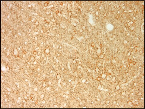
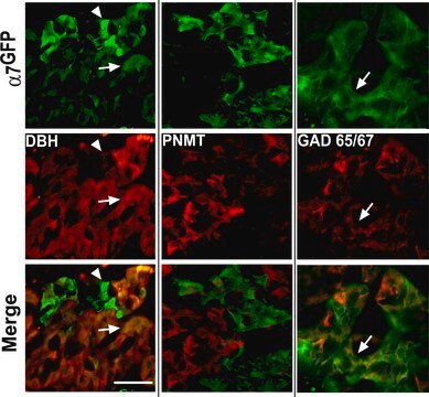
Immunohystochemical Staining Procedures

The following procedure was developed to localize GAD in rat brain sections of cerebellum. Perform all steps at room temperature unless otherwise indicated. Where normal serum is indicated, use normal serum from the same species as the source of the secondary antibody.This procedure represents suggested guidelines for the use of anti-GAD. Fixation regimen, antibody concentrations, and incubation conditions for a given experimental system should be determined empirically.

1. Perfuse rats with 100 mM phosphate buffer, pH 7.4, containing 1% paraformaldehyde, 0.34% L-lysine, and 0.05% sodium m-periodate (1% PLP).

2. Postfix brains in 1% PLP for 1-2 hours. Longer fixation times may reduce labeling intensity.

3. Transfer brains to 100 mM phosphate buffer containing 30% sucrose, and gently agitate on a shaker platform at +4°C for 48-60 hours.

4. Using a sliding microtome, cut 30 mm sections of frozen cerebellum. As the sections are cut, collect them in a vial of cold 100 mM phosphate buffer.

5. Incubate sections in phosphate-buffered saline (PBS) containing 1.5% normal serum and 0.2% TritonX-100 for 30 minutes.

6. On a shaker platform, incubate sections with anti-GAD (diluted in PBS containing 1.5% normal serum and 0.2% Triton X-100 to a final antibody concentration of 1 mg/ml) for 12-36 hours at +4°C.

7. On a shaker platform, rinse sections eight times, 10-15 minutes per rinse, in PBS.

8. Detect with a standard secondary antibody detection system (Hsu et al., 1981; Falini & Taylor, 1983; Harlow & Lane, 1988; Taylor, 1978).

9. Mount sections, dehydrate, and apply coverslips.
Research Category
Neuroscience
Research Sub Category
Neurotransmitters & Receptors

Descrição-alvo

65 kDa

forma física

Ammonium sulfate precipitation and DEAE-cellulose chromatography
Format: Purified
Lyophilized. Dissolve contents of vial in 100 µL of sterile, distilled water. This results in a final antibody concentration of 1 mg/ml in 10 mM potassium phosphate, 70 mM sodium chloride, pH 7.4 containing no preservatives.

Armazenamento e estabilidade

Maintain for 1 year at -20°C from date of shipment. Aliquot to avoid repeated freezing and thawing. For maximum recovery of product, centrifuge the original vial after thawing and prior to removing the cap.

Nota de análise

Control
Brain tissue

Outras notas

Concentration: Please refer to the Certificate of Analysis for the lot-specific concentration.

Informações legais

CHEMICON is a registered trademark of Merck KGaA, Darmstadt, Germany

Exoneração de responsabilidade

Unless otherwise stated in our catalog or other company documentation accompanying the product(s), our products are intended for research use only and are not to be used for any other purpose, which includes but is not limited to, unauthorized commercial uses, in vitro diagnostic uses, ex vivo or in vivo therapeutic uses or any type of consumption or application to humans or animals.

Não está encontrando o produto certo?  

Experimente o nosso Ferramenta de seleção de produtos.

Pictogramas

Exclamation mark

Palavra indicadora

Warning

Frases de perigo

Classificações de perigo

Acute Tox. 4 Dermal - Acute Tox. 4 Inhalation - Acute Tox. 4 Oral - Aquatic Chronic 3

Código de classe de armazenamento

13 - Non Combustible Solids

Classe de risco de água (WGK)

WGK 3

Ponto de fulgor (°F)

Not applicable

Ponto de fulgor (°C)

Not applicable


Certificados de análise (COA)

Busque Certificados de análise (COA) digitando o Número do Lote do produto. Os números de lote e remessa podem ser encontrados no rótulo de um produto após a palavra “Lot” ou “Batch”.

Já possui este produto?

Encontre a documentação dos produtos que você adquiriu recentemente na biblioteca de documentos.

Visite a Biblioteca de Documentos

Diminished neurosteroid sensitivity of synaptic inhibition and altered location of the alpha4 subunit of GABA(A) receptors in an animal model of epilepsy.
Sun, C; Mtchedlishvili, Z; Erisir, A; Kapur, J
The Journal of Neuroscience null
Time-resolved fluorometric assay for detection of autoantibodies to glutamic acid decarboxylase (GAD65).
Matti Ankelo, Annette Westerlund-Karlsson, Jorma Ilonen, Mikael Knip, Kaisa Savola et al.
Clinical Chemistry null
Galanin receptor 1 is expressed in a subpopulation of glutamatergic interneurons in the dorsal horn of the rat spinal cord.
Marc Landry, Rabia Bouali-Benazzouz, Caroline Andre, Tie Jun Sten Shi, Claire Leger et al.
The Journal of Comparative Neurology null
Anatomy of glutamic acid decarboxylase immunoreactive neurons and axons in the rat medial geniculate body.
Winer, J A and Larue, D T
The Journal of Comparative Neurology, 278, 47-68 (1988)
Distribution of alpha1, alpha4, gamma2, and delta subunits of GABAA receptors in hippocampal granule cells.
Sun, C; Sieghart, W; Kapur, J
Brain Research null

Artigos

Human iPSC neural differentiation media and protocols used to generate neural stem cells, neurons and glial cell types.

Nossa equipe de cientistas tem experiência em todas as áreas de pesquisa, incluindo Life Sciences, ciência de materiais, síntese química, cromatografia, química analítica e muitas outras.

Entre em contato com a assistência técnica