Skip to Content
MilliporeSigma
All Photos(2)

Key Documents

AB1076

Sigma-Aldrich

Anti-phospho-ILK (Ser246) Antibody

from rabbit, purified by affinity chromatography

Synonym(s):

integrin-linked kinase, 59 kDa serine/threonine-protein kinase

Sign Into View Organizational & Contract Pricing


About This Item

UNSPSC Code:
12352203
eCl@ss:
32160702
NACRES:
NA.41

biological source

rabbit

Quality Level

antibody form

affinity isolated antibody

antibody product type

primary antibodies

clone

polyclonal

purified by

affinity chromatography

species reactivity

human, rat, mouse

species reactivity (predicted by homology)

ox (based on 100% sequence homology)

technique(s)

immunofluorescence: suitable
western blot: suitable

NCBI accession no.

UniProt accession no.

shipped in

wet ice

target post-translational modification

phosphorylation (pSer246)

Gene Information

human ... ILK(3611)

General description

ILK (Integrin-Linked Kinase) is an intraceullar Ser/Thr kinase with four Ankyrin-like repeats. It is known to play pivotal roles in extracellular matrix mediated signaling. ILK is known to associate with β-Integrins in addition to many other focal adhesion associated proteins. In this context, ILK is involved cell adhesion and migration. The kinase activity appears to be reduced following integrin activation, and overexpression of p59 ILK inhibits cellular adhesion to integrin substrates. Additionally, ILK has been associated with epithelial mesechymal transition (EMT) in a TGF-β1 induced Smad dependent manner (Li, 2003). Forced expression of ILK also induced the expression of MMP-2 (matrix metalloprotease 2) and promoted cell migration and invasion (Li, 2003).

Specificity

This antibody recognizes ILK when phosphorylated at Ser246.

Immunogen

Epitope: Phosphorylated Ser246
KLH-conjugated linear peptide corresponding to human ILK phosphorylated at Ser246.

Application

Detect phospho-ILK (Ser246) using this Anti-phospho-ILK (Ser246) Antibody validated for use in WB & IF.
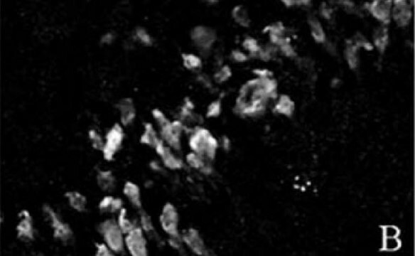
Immunofluorescence Analysis: A previous lot was used by an independent laboratory in BT549 breast cancer cells. (Dr. Shoukat Dedhar, BC Cancer Research Centre, Vancouver, BC, Canada.)
Research Category
Cell Structure
Research Sub Category
Cytoskeletal Signaling

Quality

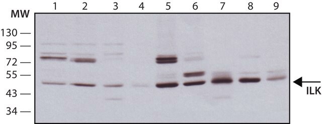
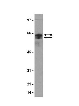
Evaluated by Western Blot in HeLa cell lysate.

Western Blot Analysis: 0.1 µg/mL of this antibody detected ILK in 10 µg of HeLa cell lysate.

Target description

~54 kDa observed

Physical form

Affinity purified
Purified rabbit polyclonal in buffer containing 0.1 M Tris-Glycine (pH 7.4), 150 mM NaCl with 0.05% sodium azide.

Storage and Stability

Stable for 1 year at 2-8°C from date of receipt.

Analysis Note

Control
HeLa cell lysate

Other Notes

Concentration: Please refer to the Certificate of Analysis for the lot-specific concentration.

Disclaimer

Unless otherwise stated in our catalog or other company documentation accompanying the product(s), our products are intended for research use only and are not to be used for any other purpose, which includes but is not limited to, unauthorized commercial uses, in vitro diagnostic uses, ex vivo or in vivo therapeutic uses or any type of consumption or application to humans or animals.

Not finding the right product?  

Try our Product Selector Tool.

Storage Class

12 - Non Combustible Liquids

wgk_germany

WGK 1

flash_point_f

Not applicable

flash_point_c

Not applicable


Certificates of Analysis (COA)

Search for Certificates of Analysis (COA) by entering the products Lot/Batch Number. Lot and Batch Numbers can be found on a product’s label following the words ‘Lot’ or ‘Batch’.

Already Own This Product?

Find documentation for the products that you have recently purchased in the Document Library.

Visit the Document Library

Matthew P Keasey et al.
Journal of cell science, 131(3) (2017-12-10)
We defined how blood-derived vitronectin (VTN) rapidly and potently activates leukemia inhibitory factor (LIF) and pro-inflammatory interleukin 6 (IL-6) in vitro and after vascular injury in the brain. Treatment with VTN (but not fibrinogen, fibronectin, laminin-111 or collagen-I) substantially increased
Victoria L Stefanelli et al.
Matrix biology : journal of the International Society for Matrix Biology, 82, 86-104 (2019-04-21)
The extracellular matrix (ECM) microenvironment is increasingly implicated in the instruction of pathologically relevant cell behaviors, from aberrant transdifferentation to invasion and beyond. Indeed, pathologic ECMs possess a panoply of alterations that provide deleterious instructions to resident cells. Here we
Jamie L Marshall et al.
The Journal of cell biology, 197(7), 1009-1027 (2012-06-27)
Utrophin is normally confined to the neuromuscular junction (NMJ) in adult muscle and partially compensates for the loss of dystrophin in mdx mice. We show that Akt signaling and utrophin levels were diminished in sarcospan (SSPN)-deficient muscle. By creating several
Jamie L Marshall et al.
Human molecular genetics, 21(20), 4378-4393 (2012-07-17)
Sarcospan (SSPN) is a core component of the major adhesion complexes in skeletal muscle, the dystrophin- and utrophin (Utr)-glycoprotein complexes (DGC and UGC). We performed a rigorous analysis of SSPN-null mice and discovered that loss of SSPN decreased DGC and

Our team of scientists has experience in all areas of research including Life Science, Material Science, Chemical Synthesis, Chromatography, Analytical and many others.

Contact Technical Service