Skip to Content
MilliporeSigma
All Photos(6)

Key Documents

L1293

Sigma-Aldrich

Anti-Lamin A (C-terminal) antibody produced in rabbit

enhanced validation

~1 mg/mL, affinity isolated antibody, buffered aqueous solution

Synonym(s):

Anti-LAMA, Anti-LMN1, Anti-LMNA, Anti-Lamin A/C

Sign Into View Organizational & Contract Pricing


About This Item

MDL number:
UNSPSC Code:
12352203
NACRES:
NA.41

biological source

rabbit

Quality Level

conjugate

unconjugated

antibody form

affinity isolated antibody

antibody product type

primary antibodies

clone

polyclonal

form

buffered aqueous solution

mol wt

antigen ~70 kDa

species reactivity

rat, human, mouse

enhanced validation

independent
Learn more about Antibody Enhanced Validation

concentration

~1 mg/mL

technique(s)

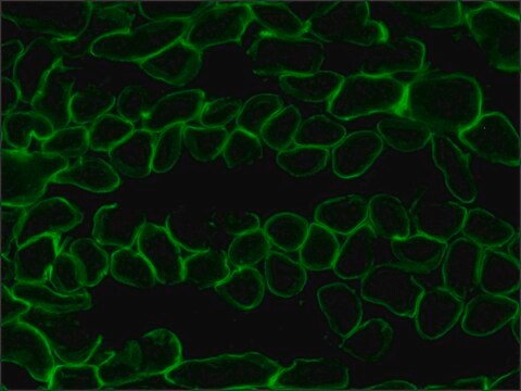
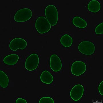
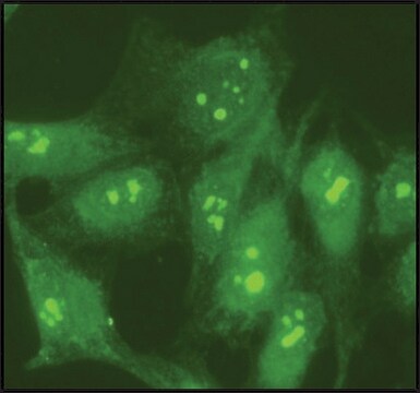
indirect immunofluorescence: 1-2 μg/mL using human HeLa, rat NRK, and mouse 3T3 cells
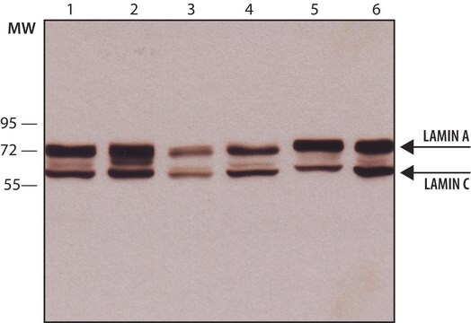
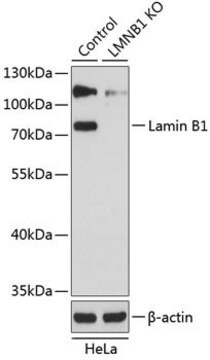
western blot: 0.1-0.2 μg/mL using human HeLa nuclear extract

UniProt accession no.

shipped in

dry ice

storage temp.

−20°C

target post-translational modification

unmodified

Gene Information

human ... LMNA(4000)
mouse ... Lmna(16905)
rat ... Lmna(60374)

General description

Lamin is a structural protein of the nuclear lamina. Lamin A is a type A lamin encoded by the LMNA gene. Lamin A contains 664 amino acids and is expressed in most somatic cells. It contains α-helical rod domain to enable assembly into filaments, a nuclear localization sequence, and a carboxy-terminal CAAX box isoprenylation sequence for nuclear membrane targeting.

Immunogen

synthetic peptide corresponding to amino acids 598-611 of human lamin A with a C-terminal added cysteine, conjugated to KLH. The corresponding sequence differs by one amino acid in rat, three amino acids in mouse, and by a gap of one amino acid in both rat and mouse lamin A.

Application

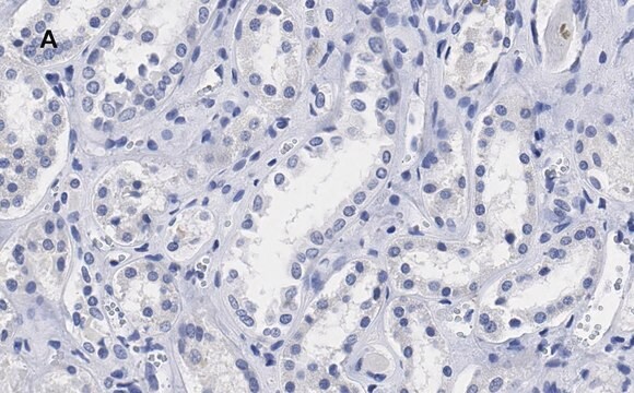
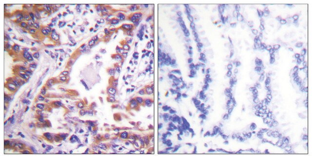
Anti-Lamin A (C-terminal) antibody is suitable for use in western blot (0.1-0.2 μg/mL) using HeLa nuclear extract. This antibody can also be used in indirect immunofluorescence (1-2 μg/mL) using HeLa cells, rat NRK and mouse 3T3 cells. Additionally, anti-Lamin A (C-terminal) antibodies are suitable for use in immunoblotting (approx. 70 kDa). Cleaved fragments of lamin A may form additional bands at 45-50 kDa.
Anti-Lamin A (C-terminal) antibody produced in rabbit has been used in:
  • western blotting
  • immunofluorescence microscopy
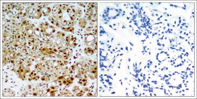
  • immunohistochemistry

Biochem/physiol Actions

Lamin A cut into a 47kDa fragment that facilitates chromatin condensation and nuclear degradation during cell death
Mutations in lamin A and C have been linked to a variety of rare human diseases including muscular dystrophy, lipodystrophy, cardiomyopathy, neuropathy, and progeroid syndromes (collectively termed laminopathies) and to premature aging (Hutchinson-Gilford progeria syndrome).

Physical form

Solution in 0.01 M phosphate buffered saline, pH 7.4, containing 15 mM sodium azide.

Disclaimer

Unless otherwise stated in our catalog or other company documentation accompanying the product(s), our products are intended for research use only and are not to be used for any other purpose, which includes but is not limited to, unauthorized commercial uses, in vitro diagnostic uses, ex vivo or in vivo therapeutic uses or any type of consumption or application to humans or animals.

Not finding the right product?  

Try our Product Selector Tool.

Storage Class

12 - Non Combustible Liquids

wgk_germany

WGK 1

flash_point_f

Not applicable

flash_point_c

Not applicable

ppe

Eyeshields, Gloves, multi-purpose combination respirator cartridge (US)


Certificates of Analysis (COA)

Search for Certificates of Analysis (COA) by entering the products Lot/Batch Number. Lot and Batch Numbers can be found on a product’s label following the words ‘Lot’ or ‘Batch’.

Already Own This Product?

Find documentation for the products that you have recently purchased in the Document Library.

Visit the Document Library

Homozygous missense mutation in the lamin A/C gene causes autosomal recessive Hutchinson-Gilford progeria syndrome
Plasilova M, et al.
Journal of medical Genetics, 41, 609-614 (2004)
Nuclear Structure and Dynamics
Cell Biology (2017)
Lamins at a glance
Ho CY and Lammerding J
Journal of Cell Science, 125(9), 2087-2093 (2012)
U Aebi et al.
Nature, 323(6088), 560-564 (1986-10-09)
The nuclear lamina, a protein meshwork lining the nucleoplasmic surface of the inner nuclear membrane, is thought to provide a framework for organizing nuclear envelope structure and an anchoring site at the nuclear periphery for interphase chromatin. In several higher
Tao Hong et al.
American journal of physiology. Endocrinology and metabolism, 300(3), E500-E507 (2010-12-16)
It has previously been known that transcription of the PGC-1α gene can be either inhibited or stimulated by p38 MAP kinase (p38 MAPK). To determine whether p38 MAPK plays an inhibitory or stimulatory role in PGC-1α gene transcription, we further

Our team of scientists has experience in all areas of research including Life Science, Material Science, Chemical Synthesis, Chromatography, Analytical and many others.

Contact Technical Service