Przejdź do zawartości
Merck
Wszystkie zdjęcia(5)

Kluczowe dokumenty

MABT831

Sigma-Aldrich

Anti-Lamin B Receptor (LBR) Antibody, clone BB2SS3F3

clone BB2SS3F3, from mouse

Synonim(y):

LMN2R, Integral nuclear envelope inner membrane protein, LBR

Zaloguj sięWyświetlanie cen organizacyjnych i kontraktowych


About This Item

Kod UNSPSC:
12352203
eCl@ss:
32160702
NACRES:
NA.41

pochodzenie biologiczne

mouse

Poziom jakości

forma przeciwciała

purified immunoglobulin

rodzaj przeciwciała

primary antibodies

klon

BB2SS3F3, monoclonal

reaktywność gatunkowa

mouse, human

opakowanie

antibody small pack of 25 μg

metody

immunocytochemistry: suitable
immunofluorescence: suitable
western blot: suitable

izotyp

IgG1κ

numer dostępu NCBI

numer dostępu UniProt

Warunki transportu

ambient

docelowa modyfikacja potranslacyjna

unmodified

informacje o genach

human ... LBR(3930)

Powiązane kategorie

Opis ogólny

Lamin-B receptor (UniProt: Q14739; also known as Integral nuclear envelope inner membrane protein, LMN2R) is encoded by the LBR gene (Gene ID: 3930) in human. Lamin-B receptor is a multi-pass membrane protein that anchors the lamina and the heterochromatin to the inner nuclear membrane. It is an integral membrane protein of the interphase nuclear envelope (NE). It contains a Tudor damain that lies within the first 62 amino acids. Its N-terminal end resides in the nucleoplasm, binding to lamin B and heterochromatin and the C-terminal end resides within the inner nuclear membrane, retreating with the endoplasmic reticulum (ER) away from condensing chromosomes during mitotic NE breakdown. During mitosis, Lamin-B receptor is retained within the retreating ER and kept out of the way of the mitotic apparatus. Lamin- B receptor is suggested to perform three vital functions in the cell: (a) contributes to post-mitotic nuclear reformation; (b) contributes to interphase NE growth; (c) contributes to compartmentalization of heterochromatin within the interphase nucleus. Lamin-B receptor is phosphorylated by Cdk1 in mitosis when the inner nuclear membrane breaks down into vesicles that dissociate from the lamina and the chromatin. It is phosphorylated by different protein kinases in interphase when the membrane is associated with these structures. Mutations in LBR gene are known to cause Pelger-Huet anomaly that is characterized by skeletal anomalies, developmental delay, and seizures. Some mutations cause Greenberg dysplasia that can lead to in utero lethality. (Ref.: Olins, AL., et al. (2010). Nucleus 1(1); 53-70).

Specyficzność

Clone BB2SS3F3 detects Lamin B receptor in human cells. It targets an epitope within the first 211 amino acids.

Immunogen

GST-tagged recombinant fragment corresponding to the first 211 amino acids from human Lamin B receptor.

Zastosowanie

Anti-Lamin B Receptor (LBR), clone BB2SS3F3, Cat. No. MABT831, is a mouse monoclonal antibody that detects Lamin B receptor and has been tested for use in Immunocytochemistry, Immunofluorescence, and Western Blotting.
Immunofluorescence Analysis: A representative lot detected Lamin B Receptor (LBR) in C57BL/6 mouse small intestine (Courtesy of Dr. Brian Burke, Institute of Medical Biology, A*STAR, Singapore).

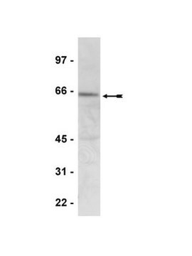
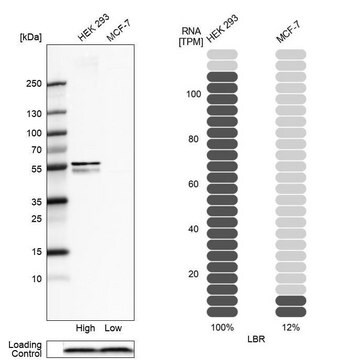
Western Blotting Analysis: A representative lot detected Lamin B Receptor (LBR) in HeLa cell lysate, mouse C2C12 cell lysate, and in adult mouse fibroblasts (MAFs) either LBR+/+ or LBR -/- (Courtesy of Dr. Brian Burke, Institute of Medical Biology, A*STAR, Singapore).

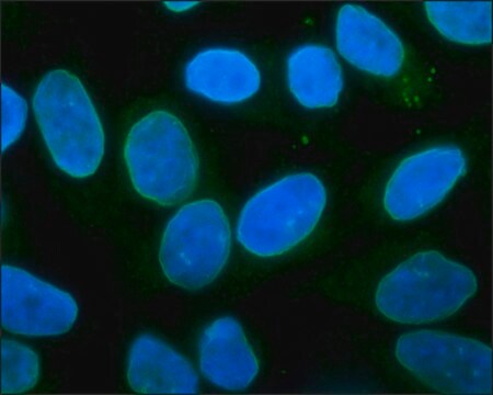
Immunocytochemistry Analysis: A 1:50 dilution from a representative lot detected Lamin B Receptor (LBR) in HeLa cells.

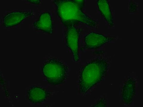
Immunocytochemistry Analysis: A representative lot detected Lamin B Receptor (LBR) in HeLa and NIE-115 cells, as well as in mouse adult fibroblasts (MAF LBR+/+ vs MAF LBR-/-) (Courtesy of Dr. Brian Burke, Institute of Medical Biology, A*STAR, Singapore).
Research Category
Cell Structure

Jakość

Evaluated by Western Blotting in mouse spleen tissue lysate.

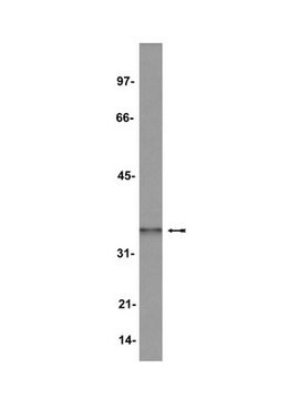
Western Blotting Analysis: 2 µg/mL of this antibody detected Lamin B Receptor (LBR) in 10 µg of mouse spleen tissue lysate.

Opis wartości docelowych

~55 kDa observed; 70.73 kDa calculated. Uncharacterized bands may be observed in some lysate(s).

Postać fizyczna

Format: Purified
Protein G purified
Purified mouse monoclonal antibody IgG1 in buffer containing 0.1 M Tris-Glycine (pH 7.4), 150 mM NaCl with 0.05% sodium azide.

Przechowywanie i stabilność

Stable for 1 year at 2-8°C from date of receipt.

Inne uwagi

Concentration: Please refer to lot specific datasheet.

Oświadczenie o zrzeczeniu się odpowiedzialności

Unless otherwise stated in our catalog or other company documentation accompanying the product(s), our products are intended for research use only and are not to be used for any other purpose, which includes but is not limited to, unauthorized commercial uses, in vitro diagnostic uses, ex vivo or in vivo therapeutic uses or any type of consumption or application to humans or animals.
Ta strona może zawierać tekst przetłumaczony maszynowo.

Nie możesz znaleźć właściwego produktu?  

Wypróbuj nasz Narzędzie selektora produktów.

Kod klasy składowania

12 - Non Combustible Liquids

Klasa zagrożenia wodnego (WGK)

WGK 1

Temperatura zapłonu (°F)

does not flash

Temperatura zapłonu (°C)

does not flash


Certyfikaty analizy (CoA)

Poszukaj Certyfikaty analizy (CoA), wpisując numer partii/serii produktów. Numery serii i partii można znaleźć na etykiecie produktu po słowach „seria” lub „partia”.

Masz już ten produkt?

Dokumenty związane z niedawno zakupionymi produktami zostały zamieszczone w Bibliotece dokumentów.

Odwiedź Bibliotekę dokumentów

Nasz zespół naukowców ma doświadczenie we wszystkich obszarach badań, w tym w naukach przyrodniczych, materiałoznawstwie, syntezie chemicznej, chromatografii, analityce i wielu innych dziedzinach.

Skontaktuj się z zespołem ds. pomocy technicznej