Przejdź do zawartości
Merck
Wszystkie zdjęcia(2)

Key Documents

MABN293

Sigma-Aldrich

Anti-Peripherin-2 Antibody, clone 6B10.1

clone 6B10.1, from mouse

Synonim(y):

Peripherin-2, Retinal degeneration slow protein, Tetraspanin-22, Tspan-22

Zaloguj sięWyświetlanie cen organizacyjnych i kontraktowych


About This Item

Kod UNSPSC:
12352203
eCl@ss:
32160702
NACRES:
NA.41

pochodzenie biologiczne

mouse

Poziom jakości

forma przeciwciała

purified immunoglobulin

rodzaj przeciwciała

primary antibodies

klon

6B10.1, monoclonal

reaktywność gatunkowa

mouse, rat, human

metody

immunohistochemistry: suitable
western blot: suitable

izotyp

IgG1κ

numer dostępu NCBI

numer dostępu UniProt

Warunki transportu

wet ice

docelowa modyfikacja potranslacyjna

unmodified

informacje o genach

human ... PRPH2(5961)

Opis ogólny

Peripherin-2 (PRPH2) is also called Retinal degeneration slow protein (RDS) and Tetraspanin-22 (Tspan-22). Peripherin-2 is a critical factor in ocular disk morphogenesis and possibly functions as an adhesion molecule helping to stabilize and compact outer segment disks, to help maintain curvature of the rim. Peripherin-2 is expressed in the photoreceptors of the retina and in the rim of rod outer segment (ROS) disks. Peripherin-2 defects are found in different retinal diseases including cone-rod dystrophy, retinitis pigmentosa, and macular degeneration.

Immunogen

Epitope: Cytoplasmic domain
GST-tagged recombinant protein corresponding to the cytoplasmic domain of human Peripherin-2.

Zastosowanie

Anti-Peripherin-2 Antibody, clone 6B10.1 is a highly specific mouse monoclonal antibody, that targets Peripherin & has been tested in western blotting & IHC.
Research Category
Neuroscience
Research Sub Category
Developmental Signaling
Western Blotting Analysis: A 1:1,000 dilution from a representative lot detected Peripherin-2 in 10 µg of human retina tissue lysate.
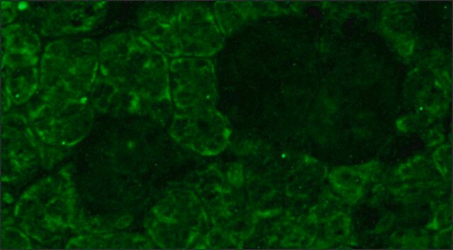
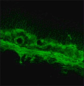
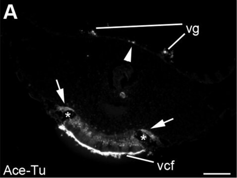
Immunohistochemistry Analysis: A 1:2,000 dilution from a representative lot detected Peripherin-2 in human retina tissue.

Jakość

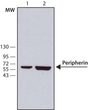
Evaluated by Western Blotting in mouse eye tissue lysate.

Western Blotting Analysis: A 1:1,000 dilution of this antibody detected Peripherin-2 in 10 µg of mouse eye tissue lysate.

Opis wartości docelowych

~35 kDa observed

Postać fizyczna

Format: Purified
Protein G Purified
Purified mouse monoclonal IgG1κ in buffer containing 0.1 M Tris-Glycine (pH 7.4), 150 mM NaCl with 0.05% sodium azide.

Przechowywanie i stabilność

Stable for 1 year at 2-8°C from date of receipt.

Oświadczenie o zrzeczeniu się odpowiedzialności

Unless otherwise stated in our catalog or other company documentation accompanying the product(s), our products are intended for research use only and are not to be used for any other purpose, which includes but is not limited to, unauthorized commercial uses, in vitro diagnostic uses, ex vivo or in vivo therapeutic uses or any type of consumption or application to humans or animals.
This page may contain text that has been machine translated.

Nie możesz znaleźć właściwego produktu?  

Wypróbuj nasz Narzędzie selektora produktów.

Kod klasy składowania

12 - Non Combustible Liquids

Klasa zagrożenia wodnego (WGK)

WGK 1

Temperatura zapłonu (°F)

Not applicable

Temperatura zapłonu (°C)

Not applicable


Certyfikaty analizy (CoA)

Poszukaj Certyfikaty analizy (CoA), wpisując numer partii/serii produktów. Numery serii i partii można znaleźć na etykiecie produktu po słowach „seria” lub „partia”.

Masz już ten produkt?

Dokumenty związane z niedawno zakupionymi produktami zostały zamieszczone w Bibliotece dokumentów.

Odwiedź Bibliotekę dokumentów

Kai Yao et al.
Nature, 560(7719), 484-488 (2018-08-17)
In zebrafish, Müller glia (MG) are a source of retinal stem cells that can replenish damaged retinal neurons and restore vision1. In mammals, however, MG do not spontaneously re-enter the cell cycle to generate a population of stem or progenitor
Katie L Bales et al.
Investigative ophthalmology & visual science, 61(10), 17-17 (2020-08-11)
To identify the role of the BBSome protein Bardet-Biedl syndrome 5 (BBS5) in photoreceptor function, protein trafficking, and structure using a congenital mutant mouse model. Bbs5-/- mice (2 and 9 months old) were used to assess retinal function and morphology.
Suguru Yamasaki et al.
iScience, 25(1), 103657-103657 (2022-01-14)
Pluripotent stem cell (PSC)-derived retinal sheet transplanted in vivo can form structured photoreceptor layers, contact with host bipolar cells, and transmit light signals to host retinas. However, a major concern is the presence of graft bipolar cells that may impede host-graft

Nasz zespół naukowców ma doświadczenie we wszystkich obszarach badań, w tym w naukach przyrodniczych, materiałoznawstwie, syntezie chemicznej, chromatografii, analityce i wielu innych dziedzinach.

Skontaktuj się z zespołem ds. pomocy technicznej