Przejdź do zawartości
Merck
Wszystkie zdjęcia(2)

Key Documents

AB5407

Sigma-Aldrich

Anti-Opsin Antibody, blue

Chemicon®, from rabbit

Synonim(y):

Anti-BCP, Anti-BOP

Zaloguj sięWyświetlanie cen organizacyjnych i kontraktowych


About This Item

Kod UNSPSC:
12352203
eCl@ss:
32160702
NACRES:
NA.41

pochodzenie biologiczne

rabbit

Poziom jakości

forma przeciwciała

purified immunoglobulin

rodzaj przeciwciała

primary antibodies

klon

polyclonal

reaktywność gatunkowa

monkey, human, mouse

producent / nazwa handlowa

Chemicon®

metody

immunohistochemistry: suitable (paraffin)

numer dostępu NCBI

numer dostępu UniProt

Warunki transportu

wet ice

docelowa modyfikacja potranslacyjna

unmodified

informacje o genach

human ... OPN1LW(5956)

Opis ogólny

The full range of color discrimination in humans is based on the presence and function of three cone photoreceptor mechanisms. Each cone type possesses a photo-sensitive pigment-protein complex consisting of 11-cis retinal and a unique opsin protein, which gives sensitivity in the short (S cone, peak sensitivity about 420nm), middle (M cone, peak sensitivity about 530nm with polymorphism; Winderckx et al., 1993; Neitz & Neitz, 1998), and long (L cone, peak sensitivity about 560nm with polymorphism; Neitz & Jacobs, 1990) wavelengths of the light spectrum. All three opsins are transmembrane proteins with seven membrane-spanning regions. Genes for the three types of cone opsins and the rod photoreceptor rhodopsin gene seem to be homologous with varying amounts of conservation. Strongest conservation is between the middle (green) and long (red) wavelength sensitive pigments on the X chromosome, suggesting a relatively recent duplication/divergence event (Nathans, 1989; Nathans et al., 1992). The S cone (blue) opsin is located on chromosome 7 and seems to have stronger conservation with rhodopsin. Cone photoreceptor distribution in humans is dominated by the M and L cone pigments.

Specyficzność

Recognizes Opsin, blue.

Immunogen

Epitope: blue
Recombinant human blue opsin.

Zastosowanie

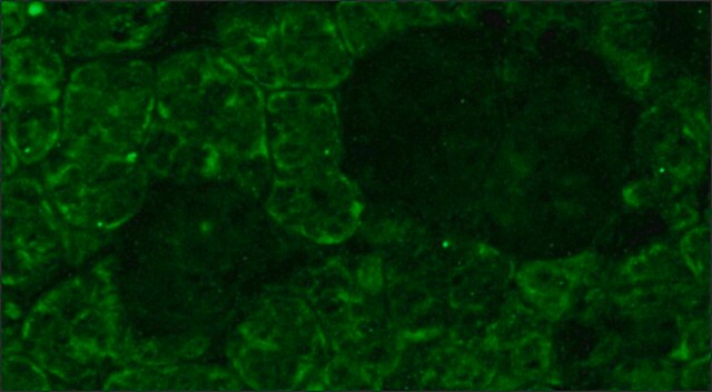
Immunohistochemistry: 1:200-1:300 on formalin-fixed, paraffin-embedded mouse retina tissue. Antigen retrieval method recommend is HIER with steam heat; other fixation and retrieval methods are untested.

Optimal working dilutions must be determined by the end user.
Research Category
Neuroscience
Research Sub Category
Sensory & PNS
This Anti-Opsin Antibody, blue is validated for use in IH(P) for the detection of Opsin.

Postać fizyczna

Format: Purified
Protein A purified
Purified immunoglobulinin PBS {0.02M phosphate, 0.25M NaCl, pH 7.6} with 0.1% sodium azide as a preservative

Przechowywanie i stabilność

Maintain for 1 year at 2–8°C from date of shipment.

Komentarz do analizy

Control
Retina

Informacje prawne

CHEMICON is a registered trademark of Merck KGaA, Darmstadt, Germany

Oświadczenie o zrzeczeniu się odpowiedzialności

Unless otherwise stated in our catalog or other company documentation accompanying the product(s), our products are intended for research use only and are not to be used for any other purpose, which includes but is not limited to, unauthorized commercial uses, in vitro diagnostic uses, ex vivo or in vivo therapeutic uses or any type of consumption or application to humans or animals.
This page may contain text that has been machine translated.

Nie możesz znaleźć właściwego produktu?  

Wypróbuj nasz Narzędzie selektora produktów.

Kod klasy składowania

12 - Non Combustible Liquids

Klasa zagrożenia wodnego (WGK)

WGK 1

Temperatura zapłonu (°F)

Not applicable

Temperatura zapłonu (°C)

Not applicable


Certyfikaty analizy (CoA)

Poszukaj Certyfikaty analizy (CoA), wpisując numer partii/serii produktów. Numery serii i partii można znaleźć na etykiecie produktu po słowach „seria” lub „partia”.

Masz już ten produkt?

Dokumenty związane z niedawno zakupionymi produktami zostały zamieszczone w Bibliotece dokumentów.

Odwiedź Bibliotekę dokumentów

Separate blue and green cone networks in the mammalian retina.
Li, Wei and DeVries, Steven H
Nature Neuroscience, 7, 751-756 (2004)
NeuroD1 regulates expression of thyroid hormone receptor 2 and cone opsins in the developing mouse retina.
Liu, H; Etter, P; Hayes, S; Jones, I; Nelson, B; Hartman, B; Forrest, D; Reh, TA
The Journal of Neuroscience null
Transcriptional profile analysis of RPGRORF15 frameshift mutation identifies novel genes associated with retinal degeneration.
Genini, S; Zangerl, B; Slavik, J; Acland, GM; Beltran, WA; Aguirre, GD
Investigative Ophthalmology & Visual Science null
The electroretinogram (ERG) of a diurnal cone-rich laboratory rodent, the Nile grass rat (Arvicanthis niloticus).
Gilmour, Gregory S, et al.
Vision Research, 48, 2723-2731 (2008)
L P Morin et al.
Neuroscience, 199, 213-224 (2011-10-12)
Four studies were performed to further clarify the contribution of rod/cone and intrinsically photoreceptive retinal ganglion cells to measures of entrainment, dark preference, light-induced locomotor suppression and photosomnolence. Wild type (WT), retinally degenerate (rd/rd), and melanopsin-less (OPN4⁻/⁻) mouse strains were

Nasz zespół naukowców ma doświadczenie we wszystkich obszarach badań, w tym w naukach przyrodniczych, materiałoznawstwie, syntezie chemicznej, chromatografii, analityce i wielu innych dziedzinach.

Skontaktuj się z zespołem ds. pomocy technicznej