
おすすめの製品


製品名
モノクロナール抗ジストロフィン マウス宿主抗体, clone MANDRA1, ascites fluid
由来生物
mouse
結合体
unconjugated
抗体製品の状態
ascites fluid
抗体製品タイプ
primary antibodies
クローン
MANDRA1, monoclonal
含みます
15 mM sodium azide
交差性
rat, human, mouse, fish
テクニック
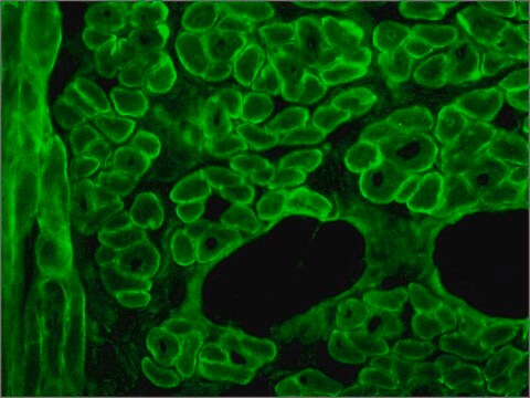
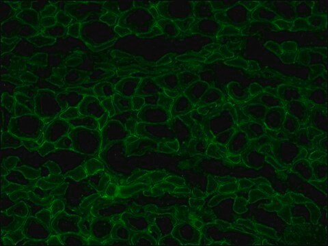
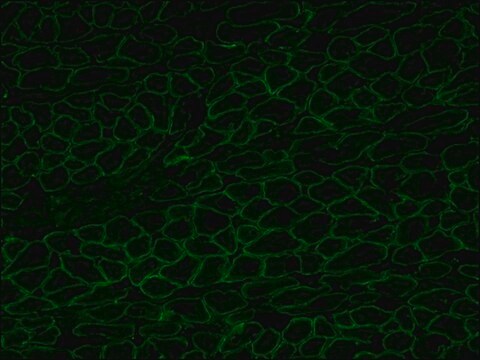
indirect immunofluorescence: 1:100 using freshly dissected and frozen human or animal muscle tissue.
microarray: suitable
アイソタイプ
IgG1
UniProtアクセッション番号
輸送温度
dry ice
保管温度
−20°C
ターゲットの翻訳後修飾
unmodified
遺伝子情報
human ... DMD(1756)
mouse ... Dmd(13405)
rat ... Dmd(24907)
詳細
Dystrophin is a muscle membrane protein (427 kDa) which is absent, reduced or altered as a result of mutation in Duchenne and Becker muscular dystrophies (DMD/BMD) or its homologue in the mouse. Severe DMD is associated with a marked dystrophin deficiency whereas patients with the milder form of DMD show less pronounced abnormalities of protein expression. Because abnormalities in the protein expression occur specifically in patients with these types of muscular dystrophy, dystrophin analysis may be used to distinguish these conditions from other neuromuscular diseases. Predictions from the sequence suggest a structural protein on the inner face of the membrane, consisting of a 25-repeat, rod-like triple-helical domain separating an N-terminal actin-binding domain from two C-terminal domains, one of which is rich in cysteine. The large size of dystrophin and its low abundance (<0.01% of the total muscle protein) are a hindrance to the isolation of intact, native protein for structure/function studies.
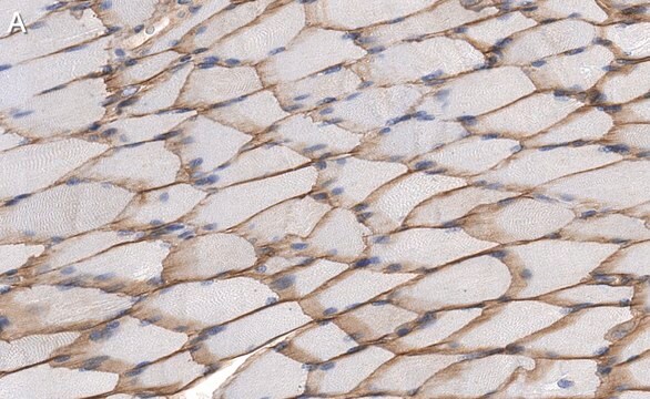
The antibody recognizes an epitope located on the 128 amino acids at the end of the C-terminal domain of the human dystrophin molecule (amino acid residues 3558-3684). Immunohistochemical staining of muscle tissue results in a clear labeling confined to the periphery (plasma membrane) of normal striated muscle fibers. By immunoblotting, the antibody stains dystrophin (427 kDa) in muscle and brain extracts. It also stains the 70-75 kDa protein known as Apo-Dystrophin-1 or Dp71.4 This is detected in the brain as well as in lymphoblastoid cells, cultures of brain astroglial and neuronal cells, liver and Hep G2 cells (human hepatoma). The epitope recognized by the antibody is sensitive to formalin fixation and paraffin embedding. The antibody exhibits a wide interspecies cross-reactivity. It is useful in ELISA and capture ELISA. The antibody is specific to dystrophin and does not react with α-actinin and utrophin, an autosomal homologue of dystrophin, also called dystrophin-related protein (DRP).
特異性
本抗体は、ヒトジストロフィン分子のC末端ドメイン末端部の128アミノ酸(アミノ酸残基3558-3684)に位置するエピト-プを認識します。筋組織の免疫組織化学染色では、正常横紋筋線維の表面(形質膜)に限局して明瞭な標識が認められています。イムノブロッティングにおいて、抗体により筋および脳抽出物のジストロフィン(427 kDa)が染色されています。また、アポジストロフィン-1あるいはDp71.4として知られている70-75 kDaのタンパク質も染色されます。これは、脳をはじめリンパ芽球様細胞、脳のアストログリアおよび神経細胞培養物、肝臓およびHep G2細胞(ヒト肝細胞腫)で検出されています。抗体により認識されるエピト-プは、ホルマリン固定、パラフィン包埋には不安定です。抗体は、広範な種間交差反応性を示します。ELISAおよび捕獲ELISAに使用できます。抗体はジストロフィンに特異的であり、α-アクチニンおよびユ-トロフィン(ジストロフィン関連タンパク質(DRP)とも呼ばれる常染色体性のジストロフィン相同体)とは反応しません。
免疫原
fusion protein containing the C-terminal amino acids of human dystrophine.
アプリケーション
Applications in which this antibody has been used successfully, and the associated peer-reviewed papers, are given below.
Immunofluorescence (1 paper)
Western Blotting (1 paper)
Immunofluorescence (1 paper)
Western Blotting (1 paper)
Mouse monoclonal clone MANDRA1 anti-Dystrophin antibody may be used for the localization of dystrophin using various immunochemical assays such as ELISA, capture ELISA, immunoblot, and immunohistochemistry. Monoclonal antibodies against defined regions of dystrophin provide a means for studying its structure and function, interactions with other proteins and the nature of the partial gene products produced in some patients carrying deletions in the dystrophin gene. The antibodies are useful in the prenatal or post-abortion diagnosis of muscular dystrophy carriers by immunohistological analyses.
ターゲットの説明
ヒトジストロフィン分子のC末端ドメイン(アミノ酸残基3558-3684)は、正常筋組織に存在しています。また、ベッカ-型筋ジストロフィ-のほぼすべてに存在していますが、デュシェンヌ型筋ジストロフィ-の症例やジストロフィ-マウス(mdx)には存在しません。
免責事項
Unless otherwise stated in our catalog or other company documentation accompanying the product(s), our products are intended for research use only and are not to be used for any other purpose, which includes but is not limited to, unauthorized commercial uses, in vitro diagnostic uses, ex vivo or in vivo therapeutic uses or any type of consumption or application to humans or animals.
適切な製品が見つかりませんか。
製品選択ツール.をお試しください
保管分類コード
13 - Non Combustible Solids
WGK
WGK 1
引火点(°F)
Not applicable
引火点(℃)
Not applicable
適用法令
試験研究用途を考慮した関連法令を主に挙げております。化学物質以外については、一部の情報のみ提供しています。 製品を安全かつ合法的に使用することは、使用者の義務です。最新情報により修正される場合があります。WEBの反映には時間を要することがあるため、適宜SDSをご参照ください。
Jan Code
D8043-.2ML:
D8043-.5ML:
D8043-BULK:
D8043-VAR:
Julia Saifetiarova et al.
Journal of neuroscience research, 95(7), 1373-1390 (2017-04-04)
Bidirectional interactions between neurons and myelinating glial cells result in formation of axonal domains along myelinated fibers. Loss of axonal domains leads to detrimental consequences on nerve structure and function, resulting in reduced conductive properties and the diminished ability to
Kazumi Zushi et al.
Journal of the Japanese Physical Therapy Association = Rigaku ryoho, 15(1), 1-8 (2012-01-01)
The purpose of this study was to investigate the effect of reloading on atrophied muscle and the time course of hypertrophy and regeneration. Forty-nine male Wistar rats were randomly assigned to groups for hindlimb suspension (HS), hindlimb suspension and reloading
Michelle Goody et al.
PLoS currents, 9 (2017-12-01)
Both genetic and infectious diseases can result in skeletal muscle degeneration, inflammation, pain, and/or weakness. Duchenne muscular dystrophy (DMD) is the most common congenital muscle disease. DMD causes progressive muscle wasting due to mutations in Dystrophin. Influenza A and B
Tomonari Awaya et al.
PloS one, 7(12), e51638-e51638 (2012-12-14)
Human embryonic stem (ES) cells and induced pluripotent stem (iPS) cells are promising sources for the cell therapy of muscle diseases and can serve as powerful experimental tools for skeletal muscle research, provided an effective method to induce skeletal muscle
Rasha Al-Khalidi et al.
Acta neuropathologica communications, 6(1), 27-27 (2018-04-13)
Duchenne muscular dystrophy (DMD) is the most common inherited muscle disorder that causes severe disability and death of young men. This disease is characterized by progressive muscle degeneration aggravated by sterile inflammation and is also associated with cognitive impairment and
ライフサイエンス、有機合成、材料科学、クロマトグラフィー、分析など、あらゆる分野の研究に経験のあるメンバーがおります。.
製品に関するお問い合わせはこちら(テクニカルサービス)