コンテンツへスキップ
Merck

366M-9

Sigma-Aldrich

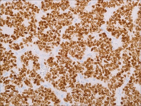
KBA.62 (KBA.62) Mouse Monoclonal Antibody

ログイン組織・契約価格を表示する


About This Item

UNSPSCコード:
12352203
NACRES:
NA.41
クローン:
KBA.62, monoclonal
application:
IHC (p)
化学種の反応性:
human
テクニック:
immunohistochemistry (formalin-fixed, paraffin-embedded sections): 1:25-1:100
citations:
4

由来生物

mouse

品質水準

100
500

結合体

unconjugated

抗体製品の状態

culture supernatant

抗体製品タイプ

primary antibodies

クローン

KBA.62, monoclonal

詳細

For In Vitro Diagnostic Use in Select Regions (See Chart)

フォーム

buffered aqueous solution

化学種の反応性

human

包装

vial of 0.1 mL concentrate (366M-94)
vial of 0.5 mL concentrate (366M-95)
bottle of 1.0 mL predilute (366M-97)
vial of 1.0 mL concentrate (366M-96)
bottle of 7.0 mL predilute (366M-98)

メーカー/製品名

Cell Marque

テクニック

immunohistochemistry (formalin-fixed, paraffin-embedded sections): 1:25-1:100

アイソタイプ

IgG1

コントロール

melanoma

輸送温度

wet ice

保管温度

2-8°C

視覚化

membranous

詳細

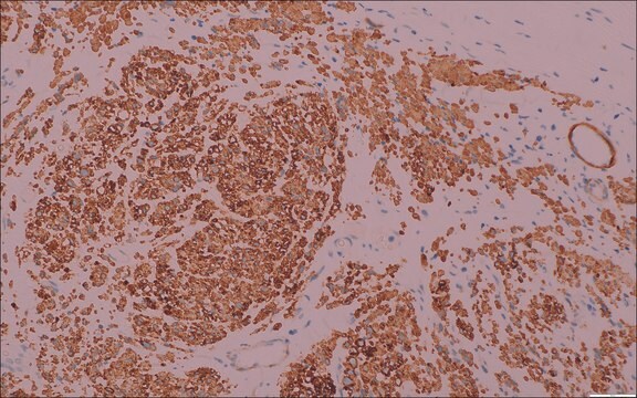
Anti-KBA.62 (Melanoma Associated Antigen) is a novel anti-melanoma antibody. Studies thus far have shown a similar sensitivity to melanocytic proliferations as that seen with S-100 protein staining, which is somewhat higher than that seen with anti-HMB-45. This has been confirmed by one study on a series of 215 sentinel lymph nodes. Moreover, anti-KBA.62 identified 6 patients (3%) who had confirmed sentinel lymph node metastasis but stained negative using anti-HMB-45. In this setting, the resolution appears to be higher than S-100 protein in that the staining pattern (membranous) is quite distinct. Interestingly, most cases of desmoplastic and spindle cell melanomas show strongly positive results with anti-KBA.62, unlike that seen with other melanocyte markers. It should be noted that anti-KBA.62 will label occasional endothelial cells which can serve as an internal positive control. A small percentage of well-differentiated squamous cell carcinomas of the skin (and lung) have also been noted to stain with this antibody; however, the poorly-differentiated forms of carcinoma do not, thus resolving a greater practical problem in differential diagnosis. Anti-KBA.62 is a useful additional marker for melanoma, specifically in desmoplastic/spindle cell cases and in the context of micrometastasis in sentinel lymph node.

関連事項

KBA.62 Positive Control Slides, Product No. 366S, are available for immunohistochemistry (formalin-fixed, paraffin-embedded sections).

物理的形状

Solution in Tris Buffer, pH 7.3-7.7, with 1% BSA and <0.1% Sodium Azide

調製ノート

Download the IFU specific to your product lot and formatNote: This requires a keycode which can be found on your packaging or product label.

その他情報

For Technical Service please contact: 800-665-7284 or email: service@cellmarque.com

法的情報

Cell Marque is a trademark of Merck KGaA, Darmstadt, Germany

適切な製品が見つかりませんか。  

製品選択ツール.をお試しください


最新バージョンのいずれかを選択してください:

試験成績書(COA)

Lot/Batch Number

申し訳ございませんが、現在この製品のCOAをオンラインで入手できません。

サポートが必要な場合は、お問い合わせください カスタマーサポート

以前この製品を購入いただいたことがある場合

文書ライブラリで、最近購入した製品の文書を検索できます。

文書ライブラリにアクセスする

P Kocan et al.
Ceskoslovenska patologie, 40(2), 50-56 (2004-07-06)
In certain primary and metastatic malignant melanomas diagnostic problems may arise due to their cytologic features and/or absence of synthesis of melanin. As the "classic" combination of S-100 protein and HMB-45 may occasionally fail to stain cells of malignant melanoma
Cécile Pagès et al.
Human pathology, 39(8), 1136-1142 (2008-05-23)
We previously described a novel antimelanoma antibody, designated KBA.62. However, review of the literature showed that only a few studies have reported on this antibody. We report our experience in the diagnosis of melanoma using KBA.62 and its value in
E Cohen-Knafo et al.
Journal of clinical pathology, 48(9), 826-831 (1995-09-01)
AIMS--To generate new monoclonal antibodies directed against melanoma associated antigens using a new melanoma cell line, KAL. METHODS--The melanoma cell line was established in culture from a lymph node metastasis of malignant melanoma. Normal Balb/c mice were immunised with KAL
O Kaufmann et al.
Modern pathology : an official journal of the United States and Canadian Academy of Pathology, Inc, 11(8), 740-746 (1998-08-28)
The authors retrospectively tested the potential value of paraffin-reactive monoclonal antibodies (A103 against melan-A, T311 against tyrosinase) and antibody KBA62 as immunohistochemical markers for amelanotic metastatic melanomas. The study cases included 72 amelanotic metastases of known cutaneous melanomas, 59 poorly

資料

Immunohistochemistry (IHC) techniques and applications have greatly improved, dermatopathology is still largely based on H&E stained slides.This paper outlines ways in which IHC antibodies can be utilized for dermatopathology.

ライフサイエンス、有機合成、材料科学、クロマトグラフィー、分析など、あらゆる分野の研究に経験のあるメンバーがおります。.

製品に関するお問い合わせはこちら(テクニカルサービス)