
MINI26
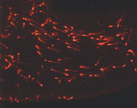
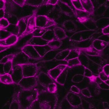
PKH26 Red Fluorescent Cell Linker Mini Kit for General Cell Membrane Labeling
Distributed for Phanos Technologies
Sinônimo(s):
Red PKH membrane label kit
About This Item
Produtos recomendados


embalagem
pkg of 1 kit
fabricante/nome comercial
Distributed for Phanos Technologies
condição de armazenamento
protect from light
técnica(s)
flow cytometry: suitable
fluorescência
λex 551 nm; λem 567 nm (PKH26 dye)
aplicação(ões)
cell analysis
detection
método de detecção
fluorometric
Condições de expedição
ambient
temperatura de armazenamento
room temp
Descrição geral
Aplicação
- labelling microglial, natural killer cells and preadipocytes
- tracking of viable transplanted adipose-derived stem cells
- in vitro proliferation studies of bone marrow stem cells
- general cell membrane labeling, including for in vitro cell labeling and long term in vivo cell tracking.
Ligação
Informações legais
Somente componentes do kit
- Diluent C 10 mL
- PKH26 Cell Linker in ethanol .1 mL


Palavra indicadora
Danger
Frases de perigo
Declarações de precaução
Classificações de perigo
Eye Irrit. 2 - Flam. Liq. 2
Código de classe de armazenamento
3 - Flammable liquids
Ponto de fulgor (°F)
57.2 °F - closed cup
Ponto de fulgor (°C)
14 °C - closed cup
Escolha uma das versões mais recentes:
Já possui este produto?
Encontre a documentação dos produtos que você adquiriu recentemente na biblioteca de documentos.
Os clientes também visualizaram


Artigos
PKH dyes are easy to use and achieve stable, uniform, and reproducible fluorescent labeling of live cells. PKH dyes are non-toxic membrane stains which produce high signal to noise ratio.
Lipophilic cell tracking dyes enable cancer biologists to track tumor and immune cell functions both in vitro and in vivo. Read the article to choose a right membrane dye kit for cell tracking and proliferation monitoring.
Optimal staining is a key component for studying tumorigenesis and progression. Learn useful tips and techniques for dye applications, including examples from recent studies.
A video about how you can use fluorescent cell tracking dyes in combination with flow and image cytometry to study interactions and fates of different cell types in vitro and in vivo.
Nossa equipe de cientistas tem experiência em todas as áreas de pesquisa, incluindo Life Sciences, ciência de materiais, síntese química, cromatografia, química analítica e muitas outras.
Entre em contato com a assistência técnica