Pular para o conteúdo
Merck
Todas as fotos(1)

Documentos Principais

C5867

Sigma-Aldrich

Anti-Ciliated Cell Marker antibody, Mouse monoclonal

clone LhS 28, purified from hybridoma cell culture

Faça loginpara ver os preços organizacionais e de contrato


About This Item

Número MDL:
Código UNSPSC:
12352203
NACRES:
NA.41

fonte biológica

mouse

Nível de qualidade

conjugado

unconjugated

forma do anticorpo

purified immunoglobulin

tipo de produto de anticorpo

primary antibodies

clone

LhS 28, monoclonal

Formulário

buffered aqueous solution

peso molecular

antigen ~45 kDa

reatividade de espécies

human, hamster

concentração

~2 mg/mL

técnica(s)

electron microscopy: suitable
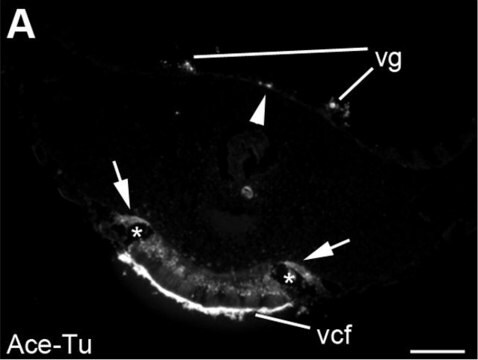
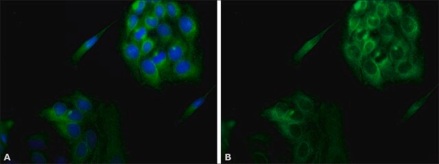
immunocytochemistry: 1-2 μg/mL using BHK a21 cells
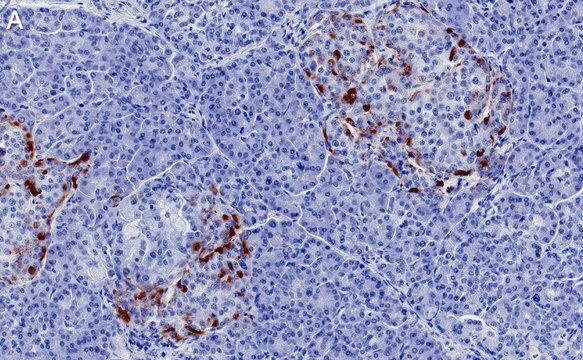
immunohistochemistry: suitable
microarray: suitable
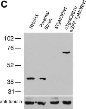
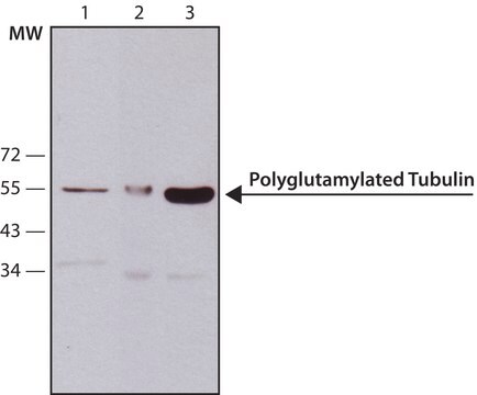
western blot: suitable

Isotipo

IgG1

Condições de expedição

dry ice

temperatura de armazenamento

−20°C

modificação pós-traducional do alvo

unmodified

Descrição geral

Anti-Ciliated Cell Marker antibody, Mouse monoclonal (mouse IgG1 isotype) is derived from the hybridoma LhS 28 produced by the fusion of mouse myeloma cells (Sp2/0 cells) and splenocytes from BALB/c mice.

Especificidade

Anti-Ciliated Cell Marker antibody, Mouse monoclonal recognizes an antigen in human and hamster ciliated cells.

Imunogênio

BHK a21 cytoskeletal preparation.

Aplicação

Anti-Ciliated Cell Marker antibody, Mouse monoclonal may be used in:
  • immunoelectron microscopy
  • immunoblotting
  • immunohistochemistry
  • immunocytochemistry

May be used in the detection of ciliated cells and for investigating mechanisms of ciliogenesis.

Ações bioquímicas/fisiológicas

The mouse hybridoma LhS 28 secretes a monoclonal antibody that recognizes the antigen 44-45 kDa protein associated with the ciliary basal bodies. The antigen recognized by LhS 28 is present in the 9+0 microtubule arrays in the basal body structure of the cilium. It serves as an unequivocal marker for in situ detection and in vitro studies of human ciliated epithelial cells. LhS28 is a potential marker for tubal metaplasia identification and may be useful in recognizing low grade cervical glandular intraepithelial neoplasia. It is effective in detecting ciliogenesis and ciliated cells associated with normal and pathological human specimens

forma física

Solution in 0.01 M phosphate buffered saline, pH 7.4, containing 15 mM sodium azide.

Armazenamento e estabilidade

For continuous use, store at 2–8 °C for up to one month. For extended storage, freeze in working aliquots. Repeated freezing and thawing, or storage in “frost-free” freezers, is not recommended. If slight turbidity occurs upon prolonged storage, clarify the solution by centrifugation before use. Working dilution samples should be discarded if not used within 12 hours.

Exoneração de responsabilidade

Unless otherwise stated in our catalog or other company documentation accompanying the product(s), our products are intended for research use only and are not to be used for any other purpose, which includes but is not limited to, unauthorized commercial uses, in vitro diagnostic uses, ex vivo or in vivo therapeutic uses or any type of consumption or application to humans or animals.

Não está encontrando o produto certo?  

Experimente o nosso Ferramenta de seleção de produtos.

Código de classe de armazenamento

10 - Combustible liquids

Classe de risco de água (WGK)

nwg

Ponto de fulgor (°F)

Not applicable

Ponto de fulgor (°C)

Not applicable


Escolha uma das versões mais recentes:

Certificados de análise (COA)

Lot/Batch Number

Não está vendo a versão correta?

Se precisar de uma versão específica, você pode procurar um certificado específico pelo número do lote ou da remessa.

Já possui este produto?

Encontre a documentação dos produtos que você adquiriu recentemente na biblioteca de documentos.

Visite a Biblioteca de Documentos

M T Comer et al.
Journal of clinical pathology, 52(5), 355-357 (1999-11-24)
The assessment of neoplastic disease in gynaecological histopathology can be complicated by the high incidence of metaplasia seen in tissues of the female genital tract. There is a need to identify specific tissue markers which can be applied in routine

Nossa equipe de cientistas tem experiência em todas as áreas de pesquisa, incluindo Life Sciences, ciência de materiais, síntese química, cromatografia, química analítica e muitas outras.

Entre em contato com a assistência técnica