18373
Atto 647N NHS ester
BioReagent, suitable for fluorescence, ≥90% (HPLC)
Faça loginpara ver os preços organizacionais e de contrato
About This Item
Produtos recomendados
linha de produto
BioReagent
Nível de qualidade
Ensaio
≥90% (HPLC)
≥90% (degree of coupling)
Formulário
solid
fabricante/nome comercial
ATTO-TEC GmbH
λ
in ethanol (with 0.1% trifluoroacetic acid)
Absorção UV
λ: 641-647 nm Amax
adequação
suitable for fluorescence
temperatura de armazenamento
−20°C
Categorias relacionadas
Descrição geral
Atto 647N is a superior red-emitting fluorescence dye with a strong absorption, excellent fluorescence quantum yield (65%), high photostability, excellent ozone resistance, good solubility, and very little triplet formation.
Aplicação
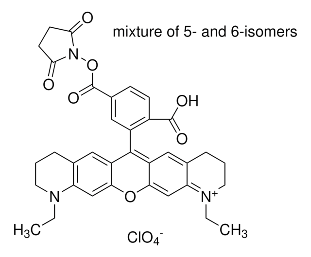
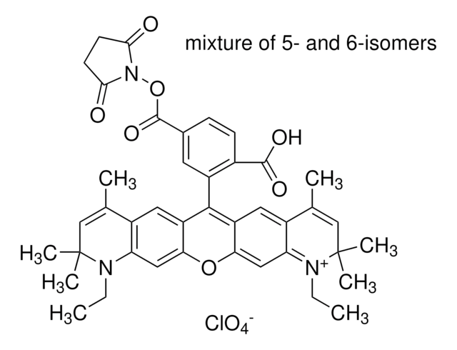
Atto 647N belongs to a new generation of fluorescent labels for the red spectral region. The dye is designed for application in the area of life science, e.g. labeling of DNA, RNA or proteins. Characteristic features of the label are strong absorption, excellent fluorescence quantum yield, high photostability, excellent ozone resistance, good solubility, and very little triplet formation. Atto 647N is a cationic dye. The active ester of Atto 647N fluorescence dye reacts with amino groups under mild conditions. After coupling to a substrate the dye carries a net electrical charge of +1. In common with most Atto-labels, absorption and fluorescence are independent of pH in the range of 2 to 11, used in typical applications. As supplied Atto 647N consists of a mixture of two isomers with practically identical absorption and fluorescence properties.
Informações legais
This product is for Research use only. In case of intended commercialization, please contact the IP-holder (ATTO-TEC GmbH, Germany) for licensing.
produto relacionado
Nº do produto
Descrição
Preços
Código de classe de armazenamento
11 - Combustible Solids
Classe de risco de água (WGK)
WGK 3
Equipamento de proteção individual
Eyeshields, Gloves, type N95 (US)
Escolha uma das versões mais recentes:
Já possui este produto?
Encontre a documentação dos produtos que você adquiriu recentemente na biblioteca de documentos.
Os clientes também visualizaram
Jialei Tang et al.
Scientific reports, 7(1), 10945-10945 (2017-09-10)
We report a simple single-molecule fluorescence imaging method that increases the temporal resolution of any type of array detector by >5-fold with full field-of-view. We spread single-molecule spots to adjacent pixels by rotating a mirror in the detection path during
Ioannis Sgouralis et al.
The journal of physical chemistry. B, 123(3), 675-688 (2018-12-21)
We develop a Bayesian nonparametric framework to analyze single molecule FRET (smFRET) data. This framework, a variation on infinite hidden Markov models, goes beyond traditional hidden Markov analysis, which already treats photon shot noise, in three critical ways: (1) it
Thorben Cordes et al.
Physical chemistry chemical physics : PCCP, 13(14), 6699-6709 (2011-02-12)
Modern fluorescence microscopy applications go along with increasing demands for the employed fluorescent dyes. In this work, we compared antifading formulae utilizing a recently developed reducing and oxidizing system (ROXS) with commercial antifading agents. To systematically test fluorophore performance in
Jennifer Z Yao et al.
Journal of the American Chemical Society, 134(8), 3720-3728 (2012-01-14)
Methods for targeting of small molecules to cellular proteins can allow imaging with fluorophores that are smaller, brighter, and more photostable than fluorescent proteins. Previously, we reported targeting of the blue fluorophore coumarin to cellular proteins fused to a 13-amino
Volker Westphal et al.
Science (New York, N.Y.), 320(5873), 246-249 (2008-02-23)
We present video-rate (28 frames per second) far-field optical imaging with a focal spot size of 62 nanometers in living cells. Fluorescently labeled synaptic vesicles inside the axons of cultured neurons were recorded with stimulated emission depletion (STED) microscopy in
Nossa equipe de cientistas tem experiência em todas as áreas de pesquisa, incluindo Life Sciences, ciência de materiais, síntese química, cromatografia, química analítica e muitas outras.
Entre em contato com a assistência técnica


