
MABF291
Anti-S100A8/S100A9 Antibody, clone 5.5
clone 5.5, from mouse
Sinônimo(s):
Protein S100-A9, Calgranulin-B, Calprotectin L1H subunit, Leukocyte L1 complex heavy chain, Migration inhibitory factor-related protein 14, S100 calcium-binding protein A9, AMRP-14, p14, Protein S100-A8, Calgranulin-A, Calprotectin L1L subunit, Cystic fi
About This Item
Produtos recomendados
fonte biológica
mouse
Nível de qualidade
forma do anticorpo
purified immunoglobulin
tipo de produto de anticorpo
primary antibodies
clone
5.5, monoclonal
reatividade de espécies
human
técnica(s)
ELISA: suitable
flow cytometry: suitable
immunohistochemistry: suitable
immunoprecipitation (IP): suitable
western blot: suitable
Isotipo
IgG1κ
Condições de expedição
wet ice
modificação pós-traducional do alvo
unmodified
Informações sobre genes
human ... S100A8(6279) , S100A9(6280)
Descrição geral
Especificidade
Imunogênio
Aplicação
Inflammation & Immunology
Immunological Signaling
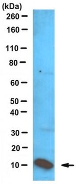
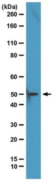
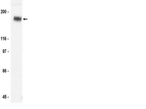
Western Blotting Analysis: A representative lot of this antibody was used to detect S100A8/S100A9 in neutrophil extracts (Hogg et al., 1989).
Immunoprecipitation Analysis: A representative lot of this antibody was used to detect S100A8/S100A9 in Human monocyte and neutrophil lysate (Edgeworth, J., et al., (1991) JBC. 266(12):7706-7713).
Immunoprecipitation Analysis: A representative lot of this antibody was used to detect S100A8/S100A9 in MRP-8 and TL-14 mutant lysate (Hessian P.A., et al., (2001) Eur. J. Biochem. 268:353-363).
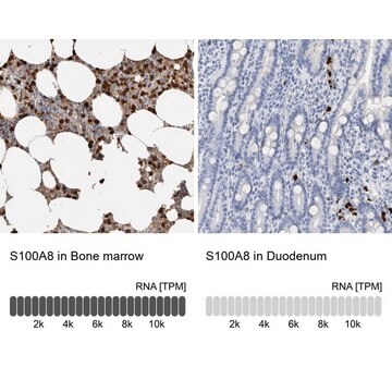
Immunohistochemistry Analysis: A representative lot of this antibody was used to detect S100A8/S100A9 in Human Bronchus tissue (Hogg, N., et al., (1989) Eur. J. Immunol. 19:1053-1061).
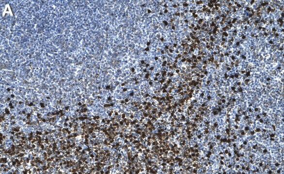
Immunohistochemistry Analysis: A representative lot of this antibody was used to detect S100A8/S100A9 in spleen and thymus tissue (Hogg, N., et al., (1989) Eur. J. Immunol. 19:1053-1061).
ELISA: A representative lot of this antibody was used to detect S100A8/S100A9 in ELISA (Ryckman, C., et al., (2003) Arthritis & Rheumatism. 48(8):2310-2320).
Qualidade
Flow Cytometry Analysis: A 1:80 dilution (0.25 µg) of this antibody detected S100A8 and/or S100A9 in 1x10E6 PBMCs.
Descrição-alvo
forma física
Armazenamento e estabilidade
Outras notas
Exoneração de responsabilidade
Não está encontrando o produto certo?
Experimente o nosso Ferramenta de seleção de produtos.
Código de classe de armazenamento
12 - Non Combustible Liquids
Classe de risco de água (WGK)
WGK 1
Ponto de fulgor (°F)
Not applicable
Ponto de fulgor (°C)
Not applicable
Certificados de análise (COA)
Busque Certificados de análise (COA) digitando o Número do Lote do produto. Os números de lote e remessa podem ser encontrados no rótulo de um produto após a palavra “Lot” ou “Batch”.
Já possui este produto?
Encontre a documentação dos produtos que você adquiriu recentemente na biblioteca de documentos.
Nossa equipe de cientistas tem experiência em todas as áreas de pesquisa, incluindo Life Sciences, ciência de materiais, síntese química, cromatografia, química analítica e muitas outras.
Entre em contato com a assistência técnica