Pular para o conteúdo
Merck
Todas as fotos(3)

Documentos Principais

MABE286

Sigma-Aldrich

Anti-Replication Protein A Antibody, clone RPA34-19

clone RPA34-19, from mouse

Sinônimo(s):

Replication protein A 32 kDa subunit, RP-A p32, Replication factor A protein 2, RF-A protein 2, Replication protein A 34 kDa subunit, RP-A p34

Faça loginpara ver os preços organizacionais e de contrato


About This Item

Código UNSPSC:
12352203
eCl@ss:
32160702
NACRES:
NA.41

fonte biológica

mouse

Nível de qualidade

forma do anticorpo

purified antibody

tipo de produto de anticorpo

primary antibodies

clone

RPA34-19, monoclonal

reatividade de espécies

human

técnica(s)

immunocytochemistry: suitable
immunohistochemistry: suitable
western blot: suitable

Isotipo

IgG1κ

nº de adesão NCBI

nº de adesão UniProt

Condições de expedição

wet ice

modificação pós-traducional do alvo

unmodified

Informações sobre genes

human ... RPA2(6118)

Descrição geral

RPA (RP-A p32) is a heterotrimeric protein complex that binds specifically to single-stranded DNA (ssDNA). It is composed of three subunits: RPA1 (70 kDa), RPA2 (32 kDa), and RPA3 (14 kDa). RPA plays multiple roles in DNA replication. At the onset of DNA replication, RPA is loaded onto chromatin, and is needed for subsequent loading of DNA polymerase and other replication proteins to initiate DNA replication. After replication begins, RPA moves with replication forks, stabilizing ssDNA and assisting in DNA synthesis. In addition to its replication function, RPA is also known to play essential roles in damage repair and recombination. The 32 kDa subunit is phosphorylated by the cdc2 family of kinases when cells enter S-phase; and by ATM, ATR, and DNA-PK proteins in response to DNA damage.

Imunogênio

Replication Protein A purified from U293 cells.

Aplicação

Anti-Replication Protein A Antibody, clone RPA34-19 is a Mouse Monoclonal Antibody for detection of Replication Protein A also known as Replication protein A 32 kDa subunit, RP-A p32 & has been validated in WB, ICC & IHC.
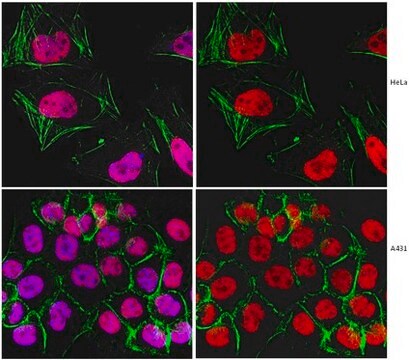
Immunocytochemistry Analysis: A 1:250 dilution from a reprsentative lot detected Replication Protein A in A431 cells.

Immunohistochemistry Analysis: A 1:5 dilution from a representative lot detected Replication Protein A in human placental chorionic villi and in human colorectal adenocarcinoma tissue.
Research Category
Epigenetics & Nuclear Function
Research Sub Category
Cell Cycle, DNA Replication & Repair

Qualidade

Evaluated by Western Blot in HeLa cell lysate.

Western Blot Analysis: A 1:2,000 dilution of this antibody detected Replication Protein A in 10 µg of HeLa cell lysate.

Descrição-alvo

~34 kDa observed. This protein has 3 isoforms: Isoform 1 (~29 kDa), Isoform 2 (~30 kDa), and Isoform 3 (~39 kDa).

forma física

Format: Purified
Protein G Purified
Purified mouse monoclonal IgG1κ in buffer containing 0.1 M Tris-Glycine (pH 7.4), 150 mM NaCl with 0.05% sodium azide.

Armazenamento e estabilidade

Stable for 1 year at 2-8°C from date of receipt.

Nota de análise

Control
HeLa cell lysate

Exoneração de responsabilidade

Unless otherwise stated in our catalog or other company documentation accompanying the product(s), our products are intended for research use only and are not to be used for any other purpose, which includes but is not limited to, unauthorized commercial uses, in vitro diagnostic uses, ex vivo or in vivo therapeutic uses or any type of consumption or application to humans or animals.

Não está encontrando o produto certo?  

Experimente o nosso Ferramenta de seleção de produtos.

Código de classe de armazenamento

12 - Non Combustible Liquids

Classe de risco de água (WGK)

WGK 1

Ponto de fulgor (°F)

Not applicable

Ponto de fulgor (°C)

Not applicable


Certificados de análise (COA)

Busque Certificados de análise (COA) digitando o Número do Lote do produto. Os números de lote e remessa podem ser encontrados no rótulo de um produto após a palavra “Lot” ou “Batch”.

Já possui este produto?

Encontre a documentação dos produtos que você adquiriu recentemente na biblioteca de documentos.

Visite a Biblioteca de Documentos

Motohiro Yamauchi et al.
DNA repair, 7(3), 405-417 (2008-02-06)
Several DNA damage checkpoint factors form nuclear foci in response to ionizing radiation (IR). Although the number of the initial foci decreases concomitantly with DNA double-strand break repair, some fraction of foci persists. To date, the physiological role of the
Janna Luessing et al.
iScience, 25(7), 104536-104536 (2022-06-28)
Abscission, the final stage of cytokinesis, occurs when the cytoplasmic canal connecting two emerging daughter cells is severed either side of a large proteinaceous structure, the midbody. Here, we expand the functions of ATR to include a cell-cycle-specific role in

Nossa equipe de cientistas tem experiência em todas as áreas de pesquisa, incluindo Life Sciences, ciência de materiais, síntese química, cromatografia, química analítica e muitas outras.

Entre em contato com a assistência técnica