Pular para o conteúdo
Merck
Todas as fotos(2)

Key Documents

MAB8151-I

Sigma-Aldrich

Anti-West Nile Virus/Kunjin Antibody, Envelope Antibody, clone 3.91D

Faça loginpara ver os preços organizacionais e de contrato


About This Item

Código UNSPSC:
12352203
NACRES:
NA.41

fonte biológica

mouse

Nível de qualidade

conjugado

unconjugated

forma do anticorpo

purified antibody

tipo de produto de anticorpo

primary antibodies

clone

3.91D, monoclonal

peso molecular

calculated mol wt 31.89 kDa
observed mol wt ~50 kDa

purificado por

using protein G

reatividade de espécies

virus

embalagem

antibody small pack of 100 μL

técnica(s)

ELISA: suitable
immunofluorescence: suitable
western blot: suitable

Isotipo

IgG3

sequência de epítopo

N-terminal half

nº de adesão de ID de proteína

nº de adesão UniProt

Condições de expedição

2-8°C

modificação pós-traducional do alvo

unmodified

Informações sobre genes

vaccinia virus ... poly> POLY(912267)

Categorias relacionadas

Descrição geral

Genome polyprotein (UniProt: P14335) is encoded by the POLY gene (Gene ID: 912267) in West Nile virus. West Nile virus (WNV) is a mosquito-borne positive-sense RNA virus of the Flavivirus genus within the Flaviviridae family. WNV genome encodes for three structural domains known as C, prM, and E and seven non-structural proteins (NS1, NS2A, NS2B, NS3, NS4A, NS4B, and NS5). These are translated as a single polypeptide, which is subsequently cleaved by viral and cellular proteases. The envelope glycoprotein (E) undergoes a conformational rearrangement triggered by low pH that results in a class II fusion event required for viral entry. The E protein (aa 291-791) contains a three-domain structure where domain I is an eight-stranded β-barrel in the center portion of the protein (aa 1-51, 134-195, and 284-297). The DII domain consists of 170 residues inserted into DI (aa 52-133 and 196-283). At the distal end of DII domain is a fusion loop (aa 98-110) and a conserved glycine-rich, hydrophobic sequence, which is important for flaviviral fusion. The third domain, DIII, is an Ig-like domain with seven β-strand and has been implicated in receptor binding. The E protein also has a single N-linked glycosylation site at Asn 154, located within the ectodomain of prM domain and is cleaved by cellular enzymes during viral particle maturation. Clone E60 specifically binds to the DII fusion loop and displays limited neutralizing activity and even at higher doses neutralization activity is shown to be between 26 and 68%. (Ref.: Martín-Acebes MA, et al. (2012). World J Virol. 1(2); 51-70).

Especificidade

Clone 3.91D is a mouse monoclonal antibody that detects West Nile virus envelope protein. It targets an epitope within the N-terminal half of polyprotein.

Imunogênio

West Nile Virus (WNV; subtype KUNV) isolate OR393.

Aplicação

Qualtiy Control Testing

Evaluated by Western Blotting with recombinant West Nile Virus envelope protein.

Western Blotting Analysis (WB): A 1:500 dilution of this antibody detected recombinant West Nile Virus envelope protein.

Tested Applications


Western Blotting Analysis: A representative lot detected West Nile Virus/Kunjin, Envelope protein in Western Blotting applications (Maeda, A., et al. (2009). Virus Res.;144(1-2):35-43; Saiyasombat, R., et al. (2014). Virol J.;11:150; Blitvich, B.J., et al. (2016). Am J Trop Med Hyg.;95(5):1185-1191).

ELISA Analysis: Various dilutions of this antibody detected recombinant West Nile Virus/Kunjin, Envelope protein.

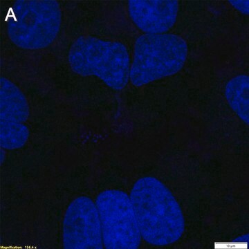
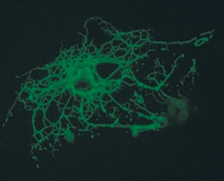
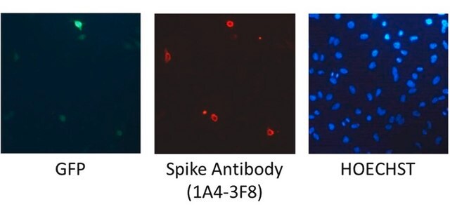
Immunofluorescence Analysis: A representative lot detected West Nile Virus/Kunjin, Envelope protein in Immunofluorescence applications (Maeda, A., et al. (2009). Virus Res.;144(1-2):35-43; Osorio, J.E., et al. (2012). Am J Trop Med Hyg.;87(3):565-72).

Note: Actual optimal working dilutions must be determined by end user as specimens, and experimental conditions may vary with the end user
Anti-West Nile Virus/Kunjin, Envelope, clone 3.91D, Cat. No. MAB8151-I, is a mouse monoclonal antibody that detects envelope protein in West Nile virus and is tested for use in ELISA, Immunofluorescence, and Western Blotting.

forma física

Purified mouse monoclonal antibody IgG3 in buffer containing 0.1 M Tris-Glycine (pH 7.4), 150 mM NaCl with 0.05% sodium azide.

Armazenamento e estabilidade

Recommend storage at +2°C to +8°C. For long term storage antibodies can be kept at -20°C. Avoid repeated freeze-thaws.

Outras notas

Concentration: Please refer to the Certificate of Analysis for the lot-specific concentration.

Exoneração de responsabilidade

Unless otherwise stated in our catalog or other company documentation accompanying the product(s), our products are intended for research use only and are not to be used for any other purpose, which includes but is not limited to, unauthorized commercial uses, in vitro diagnostic uses, ex vivo or in vivo therapeutic uses or any type of consumption or application to humans or animals.

Não está encontrando o produto certo?  

Experimente o nosso Ferramenta de seleção de produtos.

Código de classe de armazenamento

12 - Non Combustible Liquids

Classe de risco de água (WGK)

WGK 1

Ponto de fulgor (°F)

Not applicable

Ponto de fulgor (°C)

Not applicable


Certificados de análise (COA)

Busque Certificados de análise (COA) digitando o Número do Lote do produto. Os números de lote e remessa podem ser encontrados no rótulo de um produto após a palavra “Lot” ou “Batch”.

Já possui este produto?

Encontre a documentação dos produtos que você adquiriu recentemente na biblioteca de documentos.

Visite a Biblioteca de Documentos

Nossa equipe de cientistas tem experiência em todas as áreas de pesquisa, incluindo Life Sciences, ciência de materiais, síntese química, cromatografia, química analítica e muitas outras.

Entre em contato com a assistência técnica