Pular para o conteúdo
Merck
Todas as fotos(2)

Documentos Principais

ABE1957

Sigma-Aldrich

Anti-CENP-C Antibody

serum, from rabbit

Sinônimo(s):

Centromere protein C, CENP-C, CENP-C 1, Centromere autoantigen C, Centromere protein C 1, Interphase centromere complex protein 7

Faça loginpara ver os preços organizacionais e de contrato


About This Item

Código UNSPSC:
12352203
eCl@ss:
32160702

fonte biológica

rabbit

Nível de qualidade

forma do anticorpo

serum

tipo de produto de anticorpo

primary antibodies

clone

polyclonal

reatividade de espécies

human

técnica(s)

immunocytochemistry: suitable
western blot: suitable

nº de adesão NCBI

nº de adesão UniProt

Condições de expedição

ambient

modificação pós-traducional do alvo

unmodified

Informações sobre genes

human ... CENPC(1060)

Descrição geral

Centromere protein C (UniProt Q03188; also known as CENP-C, CENP-C 1, Centromere autoantigen C, Centromere protein C 1, Interphase centromere complex protein 7) is encoded by the CENPC (also known as CENPC1, ICEN7) gene (Gene ID 1060) in human. Kinetochores assemble on centromeres to bind spindle microtubules and mediate chromosome segregation during cell division. CENP-C is a subunit of constitutive centromere-associated network (CCAN) that acts as the centromere-kinetochore interface. CCAN is a large complex composed of at least 16 different centromeric proteins (CENPs). CENP-C plays an important role in kinetochore assembly downstream of CENP-A. The PEST domain in the N-terminal half of CENP-C interacts directly with the CCAN subcomplex CENP-HIKM (CENP-H, CENP-K, CENP-I, and CENP-M) and CENP-C depletion is shown to cause mislocalization of CENP-T and the CENP-HIKM complex subunits in HeLa cells.

Especificidade

This rabbit polyclonal antiserum targets the N-terminal region of human CENP-C present in both spliced isoforms reported by UniProt (Q03188). Specificity was demonstrated by a reducion of target band detection (Western blotting) and kinetochores staining (chromsome spreads fluoresence staining) following cellular CENP-C shRNA induction (Falk, S.J., et al. (2015). Science. 348(6235):699-703).

Imunogênio

Epitope: N-terminus
GST-tagged recombinant human CENP-C N-terminal fragment.

Aplicação

Anti-CENP-C Antibody, Cat. No. ABE1957, is a highly specific rabbit polyclonal antibody that targets CENP-C and has been tested in Immunocytochemistry and Western Blotting.
Immunocytochemistry Analysis: A 1:10,000 dilution from a representative lot immunostained kinetochores in chromosome spreads prepared from interphase DLD-1 human colorectal adenocarcinoma cells hypotonically swollen and fixed with 4% formaldehyde.

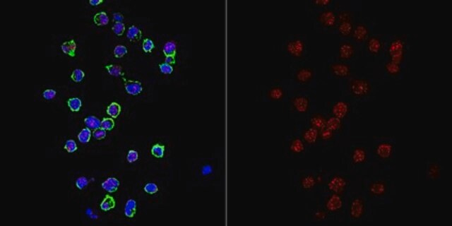
Immunocytochemistry Analysis: A representative lot was affinity purified and detected a time-dependent loss of kinetochores CENP-C immunoreactivity in 4% formaldehyde-fixed, 0.5% Triton X-100-permeabilized HeLa cells following CENP-C shRNA induction (Falk, S.J., et al. (2015). Science. 348(6235):699-703).

Immunocytochemistry Analysis: A representative lot was affinity purified and immunostained kinetochores by indirect fluorescence staining of chromosome spreads prepared from patient-derived, PD-NC4 chromosome variant harboring fibroblasts hypotonically swollen and fixed with 4% formaldehyde (Bassett, E.A., et al. (2010). J. Cell Biol. 190(2):177-185).

Western Blotting Analysis: A representative lot was affinity purified and detected a time-dependent CENP-C level in HeLa cells following CENP-C shRNA induction (Falk, S.J., et al. (2015). Science. 348(6235):699-703).
Research Category
Epigenetics & Nuclear Function

Qualidade

Evaluated by Western Blotting in DLD-1 cell lysate.

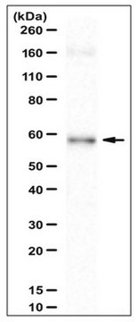
Western Blotting Analysis: A 1:5,000 dilution of this antiserum detected CENP-C in 50,000 cell equivalent of DLD-1 human colorectal adenocarcinoma cell lysate.

Descrição-alvo

~140 kDa observed. 106.8 kDa (isoform 1) and 61.54 kDa (isoform 2) calculated. The larger-than-calculated target band size is consistent with that reported in the literature (Earnshaw, W.C., et al. (1989). Chromosoma. 98(1):1-12). Uncharacterized bands may be observed in some lysate(s).

forma física

Rabbit polyclonal antibody serum with 0.05% sodium azide.
Unpurified.

Armazenamento e estabilidade

Stable for 1 year at -20°C from date of receipt.
Handling Recommendations: Upon receipt and prior to removing the cap, centrifuge the vial and gently mix the solution. Aliquot into microcentrifuge tubes and store at -20°C. Avoid repeated freeze/thaw cycles, which may damage IgG and affect product performance.

Outras notas

Concentration: Please refer to lot specific datasheet.

Exoneração de responsabilidade

Unless otherwise stated in our catalog or other company documentation accompanying the product(s), our products are intended for research use only and are not to be used for any other purpose, which includes but is not limited to, unauthorized commercial uses, in vitro diagnostic uses, ex vivo or in vivo therapeutic uses or any type of consumption or application to humans or animals.

Não está encontrando o produto certo?  

Experimente o nosso Ferramenta de seleção de produtos.

Código de classe de armazenamento

12 - Non Combustible Liquids

Classe de risco de água (WGK)

WGK 1

Ponto de fulgor (°F)

Not applicable

Ponto de fulgor (°C)

Not applicable


Certificados de análise (COA)

Busque Certificados de análise (COA) digitando o Número do Lote do produto. Os números de lote e remessa podem ser encontrados no rótulo de um produto após a palavra “Lot” ou “Batch”.

Já possui este produto?

Encontre a documentação dos produtos que você adquiriu recentemente na biblioteca de documentos.

Visite a Biblioteca de Documentos

Nossa equipe de cientistas tem experiência em todas as áreas de pesquisa, incluindo Life Sciences, ciência de materiais, síntese química, cromatografia, química analítica e muitas outras.

Entre em contato com a assistência técnica