Pular para o conteúdo
Merck
Todas as fotos(3)

Documentos

09-403

Sigma-Aldrich

Anti-TKS5 (SH3 #1) Antibody

from rabbit

Sinônimo(s):

Adaptor protein TKS5, Five SH3 domain-containing protein, SH3 and PX domains 2A, SH3 multiple domains 1, SH3 multiple domains protein 1, five SH3 domains

Faça loginpara ver os preços organizacionais e de contrato


About This Item

Código UNSPSC:
12352203
eCl@ss:
32160702
NACRES:
NA.41

fonte biológica

rabbit

Nível de qualidade

forma do anticorpo

purified antibody

tipo de produto de anticorpo

primary antibodies

clone

polyclonal

reatividade de espécies

mouse, human

técnica(s)

immunocytochemistry: suitable
immunoprecipitation (IP): suitable
western blot: suitable

nº de adesão NCBI

nº de adesão UniProt

Condições de expedição

wet ice

modificação pós-traducional do alvo

unmodified

Informações sobre genes

Descrição geral

Tks5 (previously known as Fish), is a large scaffolding protein with one amino-terminal PX domain and five SH3 domains. It is cytoplasmic in normal fibroblasts. In Src-transformed cells, Tks5 localizes to podosomes. The coexpression of Tks5 and constitutively active Src in epithelial cancer cells resulted in the appearance of podosomes/invadopodia. Cells with reduced Tks5 expression are poorly invasive through matrigel. Tks5 is expressed and localized to podosomes/invadopodia in some invasive human cancer cell lines and tumor tissue, particularly breast cancers and melanomas. Thus, Tks5 appears to be required for podosome/invadopodia formation and for invasion of some cancer cells.

Especificidade

Other homologies: Human, Canine, Equine, Rat, and Bovine (100% sequence homology), Chicken (93% sequence homology)

Imunogênio

Epitope: 166-225 aa
Recombinant Protein, aa: 166-225. Homology: Human, Dog, Horse, Rat, Cow (100%, 60/60); Chicken (93%, 56/60)

Aplicação

Anti-TKS5 (SH3 #1) Antibody detects level of TKS5 (SH3 #1) & has been published & validated for use in WB, IC & IP.
Research Category
Cell Structure
Research Sub Category
Cytoskeletal Signaling
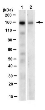
Western Blot Analysis: Antibody detected TKS5 in 20 μg of NIH-3T3 (100% confluent) lysate.

Qualidade

Routinely evaluated by Western Blot on NIH-3T3 (100% confluent) lysate.

Descrição-alvo

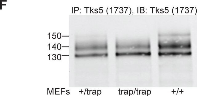
130-150 kDa Uncharacterized bands(s) may be observed in some cell lysates.

forma física

Format: Purified
Protein A purified
Purified in 0.1M Tris-Glycine (pH7.4) 150mM NaCl with 0.05% NaN3.

Armazenamento e estabilidade

Maintain at 2-8°C in undiluted aliquots for up to 1 year after date of receipt.

Nota de análise

Control
Western Blot::
NIH-3T3 (100% confluent) lysate
Immunocytochemistry:
Mouse 3T3-Src(Y527F) cells

Outras notas

Concentration: Please refer to the Certificate of Analysis for the lot-specific concentration.

Exoneração de responsabilidade

Unless otherwise stated in our catalog or other company documentation accompanying the product(s), our products are intended for research use only and are not to be used for any other purpose, which includes but is not limited to, unauthorized commercial uses, in vitro diagnostic uses, ex vivo or in vivo therapeutic uses or any type of consumption or application to humans or animals.

Not finding the right product?  

Try our Ferramenta de seleção de produtos.

Código de classe de armazenamento

12 - Non Combustible Liquids

Classe de risco de água (WGK)

WGK 1

Ponto de fulgor (°F)

Not applicable

Ponto de fulgor (°C)

Not applicable


Certificados de análise (COA)

Busque Certificados de análise (COA) digitando o Número do Lote do produto. Os números de lote e remessa podem ser encontrados no rótulo de um produto após a palavra “Lot” ou “Batch”.

Já possui este produto?

Encontre a documentação dos produtos que você adquiriu recentemente na biblioteca de documentos.

Visite a Biblioteca de Documentos

Samuel J Ghilardi et al.
Molecular biology of the cell, 32(18), 1707-1723 (2021-07-01)
Interactions between the actin cytoskeleton and the plasma membrane are important in many eukaryotic cellular processes. During these processes, actin structures deform the cell membrane outward by applying forces parallel to the fiber's major axis (as in migration) or they
Nawal Bendris et al.
Journal of cell science, 129(14), 2804-2816 (2016-06-10)
The ability of cancer cells to degrade the extracellular matrix and invade interstitial tissues contributes to their metastatic potential. We recently showed that overexpression of sorting nexin 9 (SNX9) leads to increased cell invasion and metastasis in animal models, which
Karla C Williams et al.
Oncogene, 38(19), 3598-3615 (2019-01-18)
Invadopodia are cell protrusions that mediate cancer cell extravasation but the microenvironmental cues and signaling factors that induce invadopodia formation during extravasation remain unclear. Using intravital imaging and loss of function experiments, we determined invadopodia contain receptors involved in chemotaxis
Sara K Donnelly et al.
The Journal of cell biology, 216(12), 4331-4349 (2017-10-25)
The initial step of metastasis is the local invasion of tumor cells into the surrounding tissue. Invadopodia are actin-based protrusions that mediate the matrix degradation necessary for invasion and metastasis of tumor cells. We demonstrate that Rac3 GTPase is critical

Nossa equipe de cientistas tem experiência em todas as áreas de pesquisa, incluindo Life Sciences, ciência de materiais, síntese química, cromatografia, química analítica e muitas outras.

Entre em contato com a assistência técnica