Skip to Content
MilliporeSigma
All Photos(1)

Key Documents

SAB4200237

Sigma-Aldrich

Anti-Glutathione-S-transferase (GST) antibody, Mouse monoclonal

clone 2H3-D10, purified from hybridoma cell culture

Synonym(s):

Mouse Anti-GST Tag

Sign Into View Organizational & Contract Pricing


About This Item

MDL number:
UNSPSC Code:
12352203
NACRES:
NA.41

biological source

mouse

conjugate

unconjugated

antibody form

purified immunoglobulin

antibody product type

primary antibodies

clone

2H3-D10, monoclonal

form

buffered aqueous solution

concentration

~1.0 mg/mL

technique(s)

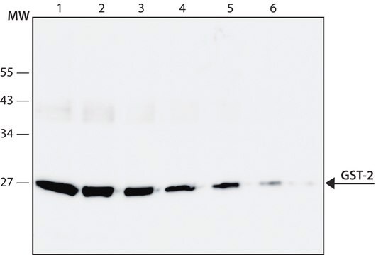
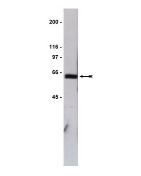
western blot: 0.2-0.4 μg/mL using detection limit for GST-fustion protein is ∼ 10ng/lane

shipped in

dry ice

storage temp.

−20°C

target post-translational modification

unmodified

General description

Anti-Glutathione-S-transferase (GST) antibody, mouse monoclonal, (mouse IgG1 isotype) is derived from the hybridoma 2H3-D10 produced by the fusion of mouse myeloma cells and splenocytes from BALB/c mice immunized with a GST-fusion protein. GST is a Phase II metabolic enzyme, expressed ubiquitously in both eukaryotes and prokaryotes. GSTs are classified in to three types based on their cellular localization, namely cytosolic GSTs, mitochondrial GSTs and microsomal GSTs.

Specificity

Monoclonal Anti- Glutathione-S-transferase (GST) recognizes GST from Schistosoma japonicum in GSTfusion proteins.

Immunogen

hybridoma 2H3-D10 produced by the fusion of mouse myeloma cells and splenocytes from BALB/c mice immunized with a GST-fusion protein

Application

Anti-Glutathione-S-transferase (GST) antibody, Mouse monoclonal has been used in immunoblotting.

Biochem/physiol Actions

Glutathione-S-Transferase (GST) is an enzyme, which catalyzes the reaction of glutathione with a wide range of electrophilic substrates and therefore, plays a role in the detoxification of potential alkylating agents.(5) GST protects the cell against oxidative stress and several toxic molecules. It facilitates the synthesis and modification of leukotrienes and prostaglandins. GST from Schistosoma japonicum, has been cloned in several expression vectors as a 27.5 kDa molecule and used as a tag for expressing proteins of interest. These proteins are thus expressed as GST-fusion proteins. Antibodies to GST are useful in identifying the successful expression of these fusion proteins.

Physical form

Solution in 0.01 M phosphate buffered saline, pH 7.4, containing 15 mM sodium azide

Storage and Stability

Store at −20 °C. For continuous use, store at 2-8 °C for up to one month. For extended storage, freeze at 20 °C in working aliquots. Repeated freezing and thawing, or storage in “frost-free” freezers, is not recommended. If slight turbidity occurs upon prolonged storage, clarify the solution by centrifugation before use. Working dilution samples should be discarded if not used within 12 hours.

Disclaimer

Unless otherwise stated in our catalog, our products are intended for research use only and are not to be used for any other purpose, which includes but is not limited to, unauthorized commercial uses, in vitro diagnostic uses, ex vivo or in vivo therapeutic uses or any type of consumption or application to humans or animals.

Not finding the right product?  

Try our Product Selector Tool.

Storage Class

10 - Combustible liquids

flash_point_f

Not applicable

flash_point_c

Not applicable


Certificates of Analysis (COA)

Search for Certificates of Analysis (COA) by entering the products Lot/Batch Number. Lot and Batch Numbers can be found on a product’s label following the words ‘Lot’ or ‘Batch’.

Already Own This Product?

Find documentation for the products that you have recently purchased in the Document Library.

Visit the Document Library

Glutathione transferases: substrates, inihibitors and pro-drugs in cancer and neurodegenerative diseases
Allocati N, et al.
Oncogenesis, 7(1), 1-15 (2018)
HnRNPK/miR-223/FBXW7 feedback cascade promotes pancreatic cancer cell growth and invasion
De He CH, et al.
Testing, 8(12), 20165-20165 (2017)
Gabriele Zaffagnini et al.
The EMBO journal, 37(5) (2018-01-19)
The removal of misfolded, ubiquitinated proteins is an essential part of the protein quality control. The ubiquitin-proteasome system (UPS) and autophagy are two interconnected pathways that mediate the degradation of such proteins. During autophagy, ubiquitinated proteins are clustered in a
Audrey W Hong et al.
Bio-protocol, 7(14) (2017-11-07)
This protocol describes a method to directly measure LATS activity by an in vitro kinase assay using YAP as a substrate.
Phospholipase Dε interacts with autophagy-related protein 8 and promotes autophagy in Arabidopsis response to nitrogen deficiency.
Shuaibing Yao et al.
The Plant journal : for cell and molecular biology, 109(6), 1519-1534 (2021-12-25)

Our team of scientists has experience in all areas of research including Life Science, Material Science, Chemical Synthesis, Chromatography, Analytical and many others.

Contact Technical Service