Skip to Content
MilliporeSigma
All Photos(6)

Key Documents

HPA008789

Sigma-Aldrich

Anti-NFATC2 antibody produced in rabbit

enhanced validation

Prestige Antibodies® Powered by Atlas Antibodies, affinity isolated antibody, buffered aqueous glycerol solution

Synonym(s):

Anti-NF-ATp antibody produced in rabbit, Anti-NFAT pre-existing subunit antibody produced in rabbit, Anti-Nuclear factor of activated T-cells, cytoplasmic 2 antibody produced in rabbit, Anti-T cell transcription factor NFAT1 antibody produced in rabbit

Sign Into View Organizational & Contract Pricing


About This Item

MDL number:
UNSPSC Code:
12352203
Human Protein Atlas Number:

biological source

rabbit

Quality Level

conjugate

unconjugated

antibody form

affinity isolated antibody

antibody product type

primary antibodies

clone

polyclonal

product line

Prestige Antibodies® Powered by Atlas Antibodies

form

buffered aqueous glycerol solution

species reactivity

human

enhanced validation

orthogonal RNAseq
Learn more about Antibody Enhanced Validation

technique(s)

immunoblotting: 0.04-0.4 μg/mL
immunofluorescence: 0.25-2 μg/mL
immunohistochemistry: 1:50-1:200

immunogen sequence

DFSILFDYEYLNPNEEEPNAHKVASPPSGPAYPDDVLDYGLKPYSPLASLSGEPPGRFGEPDRVGPQKFLSAAKPAGASGLSPRIEITPSHELIQAVGPLRMRDAGLLVEQPPLAGVAASPRFTLPV

UniProt accession no.

shipped in

wet ice

storage temp.

−20°C

target post-translational modification

unmodified

Gene Information

human ... NFATC2(4773)

General description

NFATC2 (nuclear factor of activated T-cells, cytoplasmic, calcineurin-dependent 2) is a member of the nuclear factor family called NFAT. It is expressed in peripheral T-cells, as well as cells not belonging to the immune system. It is a heavily phosphorylated protein. It was first recognized as a regulator of transcription, influencing the proliferation and migration of T-cells.

Immunogen

Nuclear factor of activated T-cells, cytoplasmic 2 recombinant protein epitope signature tag (PrEST)

Application

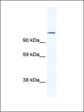
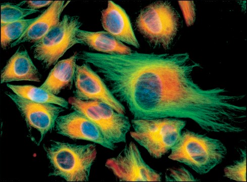
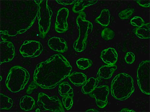
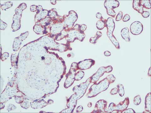
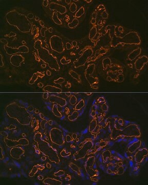
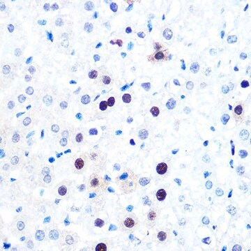
Anti-NFATC2 antibody produced in rabbit, a Prestige Antibody, is developed and validated by the Human Protein Atlas (HPA) project . Each antibody is tested by immunohistochemistry against hundreds of normal and disease tissues. These images can be viewed on the Human Protein Atlas (HPA) site by clicking on the Image Gallery link. The antibodies are also tested using immunofluorescence and western blotting. To view these protocols and other useful information about Prestige Antibodies and the HPA, visit sigma.com/prestige.
Applications in which this antibody has been used successfully, and the associated peer-reviewed papers, are given below.
Immunohistochemistry (1 paper)
Western Blotting (1 paper)

Biochem/physiol Actions

NFATC2 (nuclear factor of activated T-cells, cytoplasmic, calcineurin-dependent 2) is dephosphorylated and induced by calcineurin, which is a phosphatase. It is up-regulated in glioblastoma multiforme (GBM), where it leads to elevated expression of interleukin-13 receptor α2subunit (IL-13Rα2). In GBM, this protein facilitates the invasiveness of the tumor by activating multiple invasion-promoting genes, such as matrix metalloproteinase-7 (MMP7), MMP9 and COX2 (cyclooxygenase 2). This protein is constitutively expressed in melanoma, and its expression is suppressed on treatment with cyclosporin A, which results in death of neoplastic cells. Thus, this protein has potential as a therapeutic target in melanoma.

Features and Benefits

Prestige Antibodies® are highly characterized and extensively validated antibodies with the added benefit of all available characterization data for each target being accessible via the Human Protein Atlas portal linked just below the product name at the top of this page. The uniqueness and low cross-reactivity of the Prestige Antibodies® to other proteins are due to a thorough selection of antigen regions, affinity purification, and stringent selection. Prestige antigen controls are available for every corresponding Prestige Antibody and can be found in the linkage section.

Every Prestige Antibody is tested in the following ways:
  • IHC tissue array of 44 normal human tissues and 20 of the most common cancer type tissues.
  • Protein array of 364 human recombinant protein fragments.

Linkage

Corresponding Antigen APREST70871

Physical form

Solution in phosphate-buffered saline, pH 7.2, containing 40% glycerol and 0.02% sodium azide

Legal Information

Prestige Antibodies is a registered trademark of Merck KGaA, Darmstadt, Germany

Disclaimer

Unless otherwise stated in our catalog or other company documentation accompanying the product(s), our products are intended for research use only and are not to be used for any other purpose, which includes but is not limited to, unauthorized commercial uses, in vitro diagnostic uses, ex vivo or in vivo therapeutic uses or any type of consumption or application to humans or animals.

Not finding the right product?  

Try our Product Selector Tool.

Storage Class

10 - Combustible liquids

wgk_germany

WGK 1

flash_point_f

Not applicable

flash_point_c

Not applicable

ppe

Eyeshields, Gloves, multi-purpose combination respirator cartridge (US)


Certificates of Analysis (COA)

Search for Certificates of Analysis (COA) by entering the products Lot/Batch Number. Lot and Batch Numbers can be found on a product’s label following the words ‘Lot’ or ‘Batch’.

Already Own This Product?

Find documentation for the products that you have recently purchased in the Document Library.

Visit the Document Library

Xinxin Tie et al.
PloS one, 8(6), e66008-e66008 (2013-06-14)
Members of the nuclear factor of activated T cells (NFAT) family have been identified as regulators of oncogenic transformation in several human malignancies. A prominent member of this family, NFAT1, is associated with tumor cell survival, apoptosis, migration and invasion.
Orla Maguire et al.
Cytometry. Part A : the journal of the International Society for Analytical Cytology, 83(12), 1096-1104 (2013-10-19)
Nuclear factor of activated T cells (NFAT) is a family of transcription factors involved in regulating the immune response. The canonical NFAT pathway is calcium-dependent and upon activation, NFAT is dephosphorylated by the phosphatase, calcineurin. This results in its translocation
Matthias Weider et al.
Nature communications, 9(1), 899-899 (2018-03-04)
Oligodendrocytes produce myelin for rapid transmission and saltatory conduction of action potentials in the vertebrate central nervous system. Activation of the myelination program requires several transcription factors including Sox10, Olig2, and Nkx2.2. Functional interactions among them are poorly understood and
Valentina Perotti et al.
The Journal of investigative dermatology, 132(11), 2652-2660 (2012-06-22)
The identification of intracellular signaling pathways that promote cell proliferation and resistance to cell death may lead to the development of improved treatment for advanced melanoma. Here we show that the calcineurin/nuclear factor of activated T cells c2 (NFATc2) pathway
Qiaofang Liao et al.
Nan fang yi ke da xue xue bao = Journal of Southern Medical University, 32(6), 789-793 (2012-06-16)
To explore whether hepatitis C virus core protein (HCV C) regulates the expression of NFAT1 to participate in the progression and malignant biological behavior of intrahepatic cholangiocarcinoma cells. The recombinant plasmid pEGFP-N(3)-HCV C and the empty vector pEGFP-N(3) were cotransfected

Our team of scientists has experience in all areas of research including Life Science, Material Science, Chemical Synthesis, Chromatography, Analytical and many others.

Contact Technical Service