Skip to Content
MilliporeSigma
All Photos(3)

Key Documents

A8917

Sigma-Aldrich

Anti-Bovine IgG (whole molecule)−Peroxidase antibody produced in rabbit

IgG fraction of antiserum, buffered aqueous solution

Synonym(s):

Rabbit Anti-Bovine IgG (whole molecule)−HRP

Sign Into View Organizational & Contract Pricing


About This Item

MDL number:
UNSPSC Code:
12352203
NACRES:
NA.46

biological source

rabbit

Quality Level

conjugate

peroxidase conjugate

antibody form

IgG fraction of antiserum

antibody product type

secondary antibodies

clone

polyclonal

form

buffered aqueous solution

species reactivity

bovine

technique(s)

direct ELISA: 1:20,000
dot blot: 1:80,000
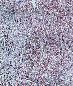
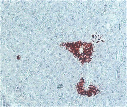
immunohistochemistry (formalin-fixed, paraffin-embedded sections): 1:1,000

shipped in

dry ice

storage temp.

−20°C

target post-translational modification

unmodified

Looking for similar products? Visit Product Comparison Guide

General description

Immunoglobulin G (IgG) contributes to 10−20% of plasma protein and is regarded as one of the most predominant serum protein. It consists of four subclasses: IgG1, IgG2, IgG3 and IgG4. The IgG structure possesses four polypeptide chains containing two identical γ heavy (H) chains and two identical κ or λ light (L) chains of 50 kDa and 25 kDa, respectively.

Immunogen

purified bovine IgG

Application

Anti-Bovine IgG (whole molecule) Peroxidase antibody produced in rabbit has been used in:
  • Dot- enzyme-linked immunosorbent assay (ELISA)
  • immunoblotting
  • immunohistology

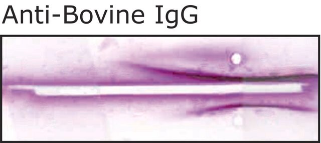
Rabbit Anti-Bovine IgG (whole molecule)-Peroxidase antibody has been used for western blot and ELISA. The antibody can also be used for dot blot (1:80,000) and immunohistochemistry (1:1,000).

Biochem/physiol Actions

Bovine IgGs are glycoprotein antibodies that regulate immune responses in herds. These antibodies inhibit TLR5 activation upon immunization with native H7 flagellin. Bovine IgG levels can be used for the detection of Johne′s disease and milk adulteration.

Physical form

Solution in 0.01 M phosphate buffered saline pH 7.4, containing 0.05% MIT

Preparation Note

Prepared by the two-step glutaraldehyde method described by Avrameas, S., et al., Scand. J. Immunol., 8, Suppl. 7, 7 (1978).

Disclaimer

Unless otherwise stated in our catalog or other company documentation accompanying the product(s), our products are intended for research use only and are not to be used for any other purpose, which includes but is not limited to, unauthorized commercial uses, in vitro diagnostic uses, ex vivo or in vivo therapeutic uses or any type of consumption or application to humans or animals.

Not finding the right product?  

Try our Product Selector Tool.

pictograms

Health hazard

signalword

Danger

hcodes

Hazard Classifications

Resp. Sens. 1 - Skin Sens. 1

Storage Class

12 - Non Combustible Liquids

wgk_germany

WGK 2

flash_point_f

Not applicable

flash_point_c

Not applicable


Certificates of Analysis (COA)

Search for Certificates of Analysis (COA) by entering the products Lot/Batch Number. Lot and Batch Numbers can be found on a product’s label following the words ‘Lot’ or ‘Batch’.

Already Own This Product?

Find documentation for the products that you have recently purchased in the Document Library.

Visit the Document Library

Investigation of antigenic specificity against Cysticercus tenuicollis cyst fluid antigen in dogs experimentally infected with Taenia hydatigena
Kara M and Douganay A
Turkish Journal of Veterinary and Animal Sciences, 29, 835-840 (2005)
Ya-mei Jin et al.
Clinical and vaccine immunology : CVI, 17(3), 476-480 (2010-01-08)
The most important animal reservoirs of Schistosoma japonicum in China are bovines. Diagnosis and control of bovine schistosomiasis is critical for reducing the prevalence of the disease. We screened defined diagnostic antigens that have the potential to increase the sensitivity
Development of Rapid Diagnostic Kit for Identification of Hanwoo (Korean Native Cattle) Brand Meat by Detecting BIO-TAG
Baek KH, et al.
Korean journal for food science of animal resources, 34(3), 339-339 (2014)
Comparison of crude and excretory/secretory antigens for the diagnosis of Fasciola hepatica in sheep by western blotting.
Kara, M. and Kircali, F.
Turkish Journal of Veterinary and Animal Sciences, 28, 943-949 (2004)
Ingrid Ieda Fernando de Souza et al.
The Journal of veterinary medical science, 81(1), 9-14 (2018-10-12)
Bovine tuberculosis (bTB) control programs generally rely on intradermal tuberculin tests for the antemortem diagnosis of Mycobacterium bovis infection in cattle, but these tests detect only a portion of the infected animals. The aim of the present study was to

Our team of scientists has experience in all areas of research including Life Science, Material Science, Chemical Synthesis, Chromatography, Analytical and many others.

Contact Technical Service