SCC179
SMA-560 Mouse Orthotopic Glioma Cell Line
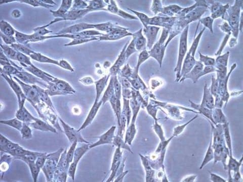
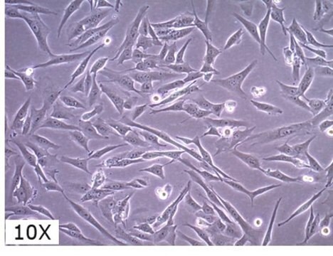
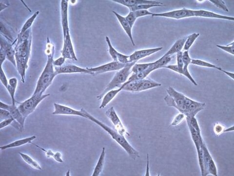
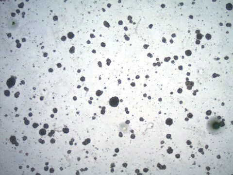
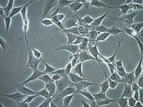
SMA-560 cell line is a well-established glioma model derived from a spontaneous murine astrocytoma.
Synonym(s):
P560, SMA 560, SMA560
Sign Into View Organizational & Contract Pricing
All Photos(1)
About This Item
UNSPSC Code:
41106514
NACRES:
NA.45
Recommended Products
technique(s)
cell culture | mammalian: suitable
General description
The SMA-560 cell line is a well-established glioma model derived from a spontaneous murine astrocytoma (2,3). SMA560 cells are highly reflective of differentiated anaplastic astrocytoma, having high expression of the differentiated astrocyte marker glial fibrillary acid protein (GFAP) and the astrocyte marker glutamine synthetase and low expression of S-100 proteins (4,5). The SMA560 cell line maintains robust tumorigenic behavior after serial passaging (2). Uniquely among established murine glioma models, SMA560 cells express the immunosuppressive protein transforming growth factor β (TGF-β) (6), making this cell line of great value for investigation of targeted cancer immunotherapies.
Source:
SMA-560 cell line was derived from a spontaneous astrocytoma passaged in syngenic VM/Dk mice (2).
Gliomas are a rare and aggressive cancer with an extremely low survival rate and high resistance to treatment (1). Glioma models that recapitulate the multiple features of the disease are important for understanding of mechanisms of glioma malignancy and development of effective therapies, especially those that address tumor-induced immunosuppression and resistance.
References:
1. N Engl J Med 2005; 352(10): 987-996.
2. Acta Neuropathol 1980; 51(1): 53-64.
3. Neurosci Lett 1982; 34(3): 315-320.
4. J Neurol Sci 1983; 62(1-3): 115-139.
5. J Neurol Neurosurg Psychiatry 1986; 49(12): 1361-1366.
6. Neurosurgery 1997; 41(6): 1365-1372.
Cell Line Description
Cancer Cells
Application
SMA-560 cell line is a well-established glioma model derived from a spontaneous murine astrocytoma.
Packaging
≥1X10⁶ cells/vial
Quality
- Each vial contains ≥ 1X10⁶ viable cells.
- Cells are tested negative for infectious diseases by a Mouse Essential CLEAR panel by Charles River Animal Diagnostic Services.
- Cells are verified to be of mouse origin and negative for inter-species contamination from rat, chinese hamster, Golden Syrian hamster, human and non-human primate (NHP) as assessed by a Contamination Clear panel by Charles River Animal Diagnostic Services
- Cells are negative for mycoplasma contamination.
Storage and Stability
Store in liquid nitrogen. The cells can be cultured for at least 10 passages after initial thawing without significantly affecting the cell marker expression and functionality.
Disclaimer
This product is intended for sale and sold solely to academic institutions for internal academic research use per the terms of the “Academic Use Agreement” as detailed in the product documentation. For information regarding any other use, please contact licensing@emdmillipore.com.
Unless otherwise stated in our catalog or other company documentation accompanying the product(s), our products are intended for research use only and are not to be used for any other purpose, which includes but is not limited to, unauthorized commercial uses, in vitro diagnostic uses, ex vivo or in vivo therapeutic uses or any type of consumption or application to humans or animals.
Storage Class
12 - Non Combustible Liquids
wgk_germany
WGK 1
flash_point_f
Not applicable
flash_point_c
Not applicable
Certificates of Analysis (COA)
Search for Certificates of Analysis (COA) by entering the products Lot/Batch Number. Lot and Batch Numbers can be found on a product’s label following the words ‘Lot’ or ‘Batch’.
Already Own This Product?
Find documentation for the products that you have recently purchased in the Document Library.
Our team of scientists has experience in all areas of research including Life Science, Material Science, Chemical Synthesis, Chromatography, Analytical and many others.
Contact Technical Service