Skip to Content
MilliporeSigma
All Photos(9)

Key Documents

MAB424

Sigma-Aldrich

Anti-PCNA Antibody, clone PC10

clone PC10, Chemicon®, from mouse

Synonym(s):

Proliferating Cell Nuclear Antigen, DNA Polymerase delta Processivity Factor

Sign Into View Organizational & Contract Pricing


About This Item

UNSPSC Code:
12352203
eCl@ss:
32160702
NACRES:
NA.41

biological source

mouse

Quality Level

antibody form

purified immunoglobulin

antibody product type

primary antibodies

clone

PC10, monoclonal

species reactivity

vertebrates, invertebrates

packaging

antibody small pack of 25 μg

manufacturer/tradename

Chemicon®

technique(s)

ELISA: suitable
flow cytometry: suitable
immunohistochemistry: suitable (paraffin)
immunoprecipitation (IP): suitable
western blot: suitable

isotype

IgG2a

NCBI accession no.

UniProt accession no.

shipped in

ambient

storage temp.

2-8°C

target post-translational modification

unmodified

Gene Information

human ... PCNA(5111)

Related Categories

General description

Proliferating cell nuclear antigen (PCNA) is a 36 kDa molecule that is highly conserved between species. PCNA was first identified as the antigen for a subpopulation of autoantibodies in patients with systemic lupus erythematosus (Miyachi, 1978; Takasaki, 1984; Ogata, 1987). It has since been determined that PCNA serves as a co-factor for DNA polymerase delta in S-phase as well as during DNA synthesis associated with mechanisms involved in DNA damage repair (Tan, 1987; Bravo, 1987). The temporal specificity of PCNA expression makes it an ideal marker for cell proliferation. PCNA begins to accumulate during the G1 phase of the cell cycle, is most abundant during the S phase, and declines during the G2/M phase (Kurki, 1988). Since the half-life of PCNA exceeds 20 hours, it may be possible to detect the protein in non-cycling cells.

Specificity

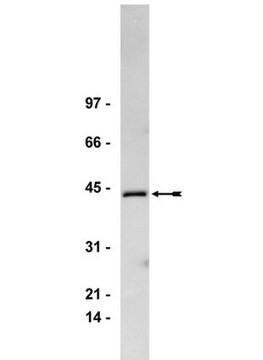
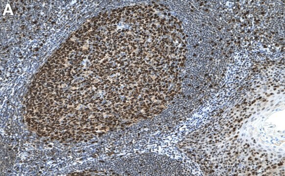
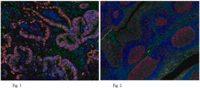
Clone PC10 recognizes PCNA from all vertebrate and insect species tested and has been used to identify transformed cells (Kurki, 1988), proliferating cells in solid tumors (Smetana, 1983), and blast cells in leukemia patients (Takasaki, 1984). Immunohistochemistry with clone PC10 has been used to study the expression of PCNA in paraffin sections of normal tissues and lymphoid neoplasms (Hall, 1990). In proliferating cells, PCNA staining pattern is predominantly nuclear. By western blot, the antibody detects a polypeptide migrating at 36 kDa with an isoelectric point of 4.8.

Immunogen

Rat PCNA made in the protein A vector pR1T2T.

Application

Research Category
Epigenetics & Nuclear Function
Research Sub Category
Cell Cycle, DNA Replication & Repair
Use Anti-PCNA Antibody, clone PC10 (mouse monoclonal antibody) validated in ELISA, FC,IHC(P), IP, WB to detect PCNA also known as Proliferating Cell Nuclear Antigen, DNA Polymerase delta Processivity Factor.
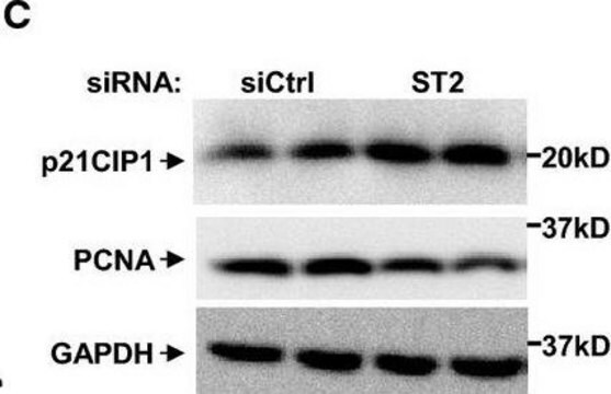
Western blot: 1:250-1:1000. Nuclear extracts are preferred over whole cell protein extracts.

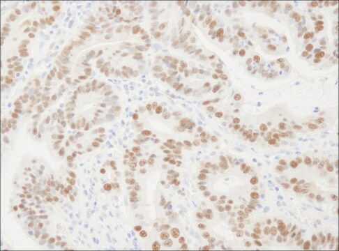
Immunohistochemistry: 1:20-1:200. Recommended for paraffin embedded tissue sections only. May be used on material fixed in a wide range of fixatives including formalin (buffered and unbuffered), methacarn and Bouin′s reagent. The time of fixation can markedly affect the intensity of PCNA immunoreactivity. Staining is reduced (and may be abolished) if sections are baked onto glass slides. Air-drying overnight on poly-L-lysine coated slides is recommended. 60 minute incubation at 25°C with standard ABC technique is recommended.

Immunoprecipitation

Indirect Flow Cytometry

Optimal working dilutions must be determined by the end user.

Quality

Tested

Target description

36 kDa

Physical form

Format: Purified
Protein A Purified mouse immunoglobulin in PBS with 0.1% sodium azide as a preservative.
Protein A purified

Storage and Stability

Maintain for 1 year at 2–8°C from date of shipment. Aliquot to avoid repeated freezing and thawing. For maximum recovery of product, centrifuge the original vial after thawing and prior to removing the cap.

Analysis Note

Control
Rat kidney, Human tonsil, lymph node tissue

Other Notes

Concentration: Please refer to the Certificate of Analysis for the lot-specific concentration.

Legal Information

CHEMICON is a registered trademark of Merck KGaA, Darmstadt, Germany

Disclaimer

Unless otherwise stated in our catalog or other company documentation accompanying the product(s), our products are intended for research use only and are not to be used for any other purpose, which includes but is not limited to, unauthorized commercial uses, in vitro diagnostic uses, ex vivo or in vivo therapeutic uses or any type of consumption or application to humans or animals.

Not finding the right product?  

Try our Product Selector Tool.

Storage Class

10 - Combustible liquids

wgk_germany

WGK 2

flash_point_f

Not applicable

flash_point_c

Not applicable


Certificates of Analysis (COA)

Search for Certificates of Analysis (COA) by entering the products Lot/Batch Number. Lot and Batch Numbers can be found on a product’s label following the words ‘Lot’ or ‘Batch’.

Already Own This Product?

Find documentation for the products that you have recently purchased in the Document Library.

Visit the Document Library

Spatial and temporal expression of RP58, a novel zinc finger transcriptional repressor, in mouse brain.
Chiaki Ohtaka-Maruyama, Akiko Miwa, Hitoshi Kawano, Masataka Kasai, Haruo Okado
The Journal of Comparative Neurology null
PCNA and Ki-67 immunoreactivity in multinucleated cells of giant cell fibroma and peripheral giant cell granuloma.
A J Mighell, P A Robinson, W J Hume
Journal of Oral Pathology & Medicine null
Stepwise activation of the ATR signaling pathway upon increasing replication stress impacts fragile site integrity.
Koundrioukoff, S; Carignon, S; Techer, H; Letessier, A; Brison, O; Debatisse, M
PLoS Genetics null
Autoantibody to the proliferating cell nuclear antigen neutralizes the activity of the auxiliary protein for DNA polymerase delta.
Tan, C K, et al.
Nucleic Acids Research, 15, 9299-9308 (1987)
Monoclonal antibodies to proliferating cell nuclear antigen (PCNA)/cyclin as probes for proliferating cells by immunofluorescence microscopy and flow cytometry.
Kurki, P, et al.
Journal of Immunological Methods, 109, 49-59 (1988)

Our team of scientists has experience in all areas of research including Life Science, Material Science, Chemical Synthesis, Chromatography, Analytical and many others.

Contact Technical Service