Skip to Content
MilliporeSigma
All Photos(1)

Key Documents

MAB1594

Sigma-Aldrich

Anti-Frataxin Antibody, exon 4, clone 1G2

ascites fluid, clone 1G2, Chemicon®

Synonym(s):

Friedreich ataxia, Friedreich ataxia protein

Sign Into View Organizational & Contract Pricing


About This Item

UNSPSC Code:
12352203
eCl@ss:
32160702
NACRES:
NA.41

biological source

mouse

Quality Level

antibody form

ascites fluid

antibody product type

primary antibodies

clone

1G2, monoclonal

species reactivity

human, rat, mouse

manufacturer/tradename

Chemicon®

technique(s)

ELISA: suitable
immunocytochemistry: suitable
immunofluorescence: suitable
western blot: suitable

isotype

IgG1κ

NCBI accession no.

UniProt accession no.

shipped in

dry ice

target post-translational modification

unmodified

Gene Information

human ... FXN(2395)
mouse ... Fxn(14297)
rat ... Fxn(499335)

General description

Frataxin is a monomeric mitochondrial protein that is believed to be involved in iron homeostasis through an unknown mechanism. Expression of frataxin is highest in tissue rich with mitochondria including liver, heart, and skeletal muscle (Campuzano, 1996; Koutnikova, 1997). Frataxin is expressed as a 30 kDa precursor (transient; 210 amino acids) that is processed within in the mitochondria in two steps catalysed by the mitochondrial processing peptidase (MPP) to yield the mature protein (Koutnikova, 1998). The first step involves cleavage of the first 41 N-terminal amino acids by MPP yielding a transient intermediate of approximately 20 kDa (aa 42-210). Further cleavage of the N-terminus of this 20 kDa intermediate by MPP results in the mature 18 kDa frataxin protein (aa 56-210). Defects in the gene encoding frataxin are implicated as the cause of Friedreich′s ataxia, an autosomal recessive, progressive degenerative disease characterized by neurodegeneration and cardiomyopathy. In the majority of cases of Friedreich′s ataxia, there is an expansion of a trinucleotide repeat in the first intron of the gene encoding frataxin resulting in a marked decrease in frataxin expression, perhaps due to the formation of some unusual yet stable DNA structure that interferes with transcription (Campuzano, 1997; Bidichandani, 1998). This reduction in frataxin expression results in the accumulation of unchelated iron in the mitochondria, inhibition of mitochondrial iron-sulfer proteins, and iron mediated oxidative stress (Foury, 1997; for review see Puccio, 2000).

Specificity

Expected to cross-react with human.
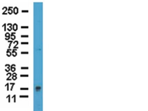
Human Frataxin. MAB1594 recognizes only isoforms of frataxin containing exon 4. On Western blots of normal human muscle, heart, cerebellum, and spinal cord extracts, MAB1594 recognizes a band migrating at approximately 18 kDa corresponding to processed frataxin (Campuzano, 1997). Slight cross reactivity with myosin may be observed by Western blot. Immunofluorescent labeling of HeLa cells with MAB1594 demonstrates that frataxin is predominantly localized in the mitochondria (Campuzano, 1997).

Immunogen

Epitope: exon 4
Full length human Frataxin fused to TrpE.

Application

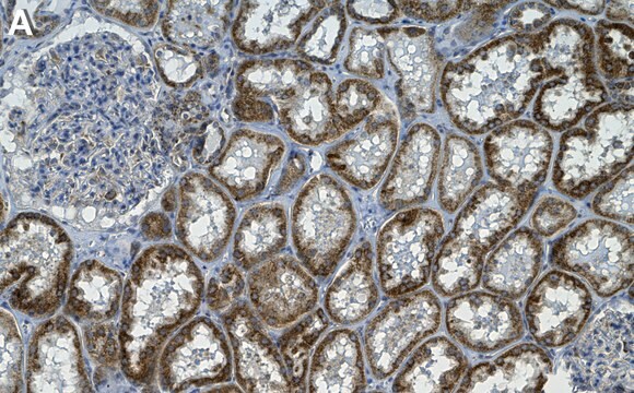
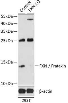
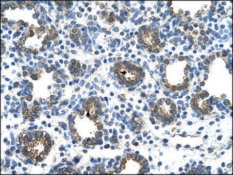
Detect Frataxin using this Anti-Frataxin Antibody, exon 4, clone 1G2 validated for use in ELISA, IC, IF & WB.
Immunofluorescence:
1:100-1:1,000. Fixation of cells in ice cold acetone or 4% paraformaldehyde is recommended. Due to the subcellular localization of frataxin in the mitochondria, cells should be permeabilized in the presence of detergent prior to incubation with primary antibody.

ELISA:
A previous lot of this antibody was used on ELISA.

Western blot (natural and recombinant protein):
1:5,000; mitochondrial preparations are recommended for consist signals (see Santos, 2001).

Optimal working dilutions must be determined by the end user.

Quality

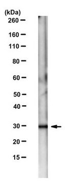
Routinely evaluated by Western Blot on PC12 lysates.

Western Blot Analysis:
1:1000 dilution of this lot detected Frataxin on 10 μg of PC12 lysates.

Target description

~ 18 kDa

Physical form

Mouse monoclonal ascites IgG1κ in buffer containing liquid with no preservatives.

Storage and Stability

Stable for 1 years at -20°C in undiluted aliquots from date of receipt.
Handling Recommendations: Upon first thaw, and prior to removing the cap, centrifuge the vial and gently mix the solution. Aliquot into microcentrifuge tubes and store at -20°C. Avoid repeated freeze/thaw cycles, which may damage IgG and affect product performance

Analysis Note

Control
Liver, heart or skeletal muscle.

Other Notes

Concentration: Please refer to the Certificate of Analysis for the lot-specific concentration.

Legal Information

CHEMICON is a registered trademark of Merck KGaA, Darmstadt, Germany

Not finding the right product?  

Try our Product Selector Tool.

Storage Class

11 - Combustible Solids

wgk_germany

WGK 1

flash_point_f

Not applicable

flash_point_c

Not applicable


Certificates of Analysis (COA)

Search for Certificates of Analysis (COA) by entering the products Lot/Batch Number. Lot and Batch Numbers can be found on a product’s label following the words ‘Lot’ or ‘Batch’.

Already Own This Product?

Find documentation for the products that you have recently purchased in the Document Library.

Visit the Document Library

Erythroid differentiation and protoporphyrin IX down-regulate frataxin expression in Friend cells: characterization of frataxin expression compared to molecules involved in iron metabolism and hemoglobinization
Becker, Erika M, et al
Blood, 99, 3813-3822 (2002)
Hannes Steinkellner et al.
Analytica chimica acta, 659(1-2), 129-132 (2010-01-28)
Friedreich's ataxia (FRDA) is an autosomal recessive neurodegenerative disease affecting 1 in 50,000 people and is caused by a GAA-trinucleotide expansion in the frataxin gene located on chromosome locus 9q13 which results in a markedly reduced expression of frataxin, a
H Koutnikova et al.
Human molecular genetics, 7(9), 1485-1489 (1998-08-13)
Frataxin is a mitochondrial protein deficient in Friedreich ataxia (FRDA) and which is associated with abnormal intramitochondrial iron handling. We identified the mitochondrial processing peptidase beta (MPPbeta) as a frataxin protein partner using the yeast two-hybrid assay. In in vitro
Infectious delivery and expression of a 135 kb human FRDA genomic DNA locus complements Friedreich's ataxia deficiency in human cells.
Gomez-Sebastian, Silvia, et al.
Molecular Therapy, 15, 248-254 (2007)
M M Santos et al.
Human molecular genetics, 10(18), 1935-1944 (2001-09-14)
Deficiency of the mitochondrial matrix protein frataxin causes Friedreich ataxia. Frataxin function is believed to be related to mitochondrial iron metabolism and free radical production. In Friedreich ataxia, loss of dorsal root ganglia neurons occurs early in life, suggesting a

Our team of scientists has experience in all areas of research including Life Science, Material Science, Chemical Synthesis, Chromatography, Analytical and many others.

Contact Technical Service