Skip to Content
MilliporeSigma
All Photos(6)

Key Documents

MAB1501R

Sigma-Aldrich

Anti-Actin Antibody, clone C4

clone C4, Chemicon®, from mouse

Synonym(s):

actin, alpha 1, skeletal muscle, alpha skeletal muscle actin, MAB1501, MAB1501X, smooth muscle actin, SMA

Sign Into View Organizational & Contract Pricing


About This Item

UNSPSC Code:
12352203
eCl@ss:
32160702
NACRES:
NA.41

biological source

mouse

Quality Level

antibody form

purified antibody

antibody product type

primary antibodies

clone

C4, monoclonal

purified by

using protein G

species reactivity (predicted by homology)

all

manufacturer/tradename

Chemicon®

technique(s)

ELISA: suitable
immunocytochemistry: suitable
immunohistochemistry (formalin-fixed, paraffin-embedded sections): suitable
western blot: suitable

isotype

IgG2bκ

NCBI accession no.

UniProt accession no.

shipped in

wet ice

target post-translational modification

unmodified

Gene Information

human ... ACTA1(58)

General description

Actins are ubiquitous eukaryotic proteins that serve as a multi-functional, basic building blocks of cytoskeletal microfilaments. They play critical roles in a wide range of cellular processes, including cell division, cell migration, chromatin remodeling, trascriptional regulation and vesicle trafficking. These funstions are attributed to their ability to form filaments, which can quickly assemble and disassemble depending upon the needs of the cell. At least six different actin types have been reported in mammals. Although actins show about 90% overall sequence homology, isoforms do not show spatial, temporal and tissue-specific expression patterns and only 50-60% homology is found in their 18 N-terminal residues. Beta and gamma-actins, also known as cytoplasmic actins, are highly conserved in higher animals and are predominantly expressed in non-muscle cells where they control cell structure. Exocytosis, and motility. They are nearly identical proteins and differ only in four amino acids at the N-terminal region. The other four actin isoforms are typically found in specific adult muscle tissue types. Alpha-cardiac and alpha-skeletal actins are expressed in striated cardiac and skeletal muscles, respectively. Alpha and gamma actins are primarily found in vascular smooth muscle and enteric smooth muscles, respectively. It has been shown that under calcium-bound conditions, beta-actin exhibits more dynamic behavior than gamma-actin with higher rates of polymerization and depolymerization. Also, beta- and gamma-actins can readily copolymerize, and the resulting filaments exhibit polymerization and depolymerization rates that vary depending on the ration of beta- to gamma-actin (Lessard, JL.,et al.(1988). Cell Motility Cytoskeleton 10(3); 349-362.

Specificity

MAB1501R is a pan-actin antibody that binds to an epitope in a highly conserved region of actin; therefore, this antibody reacts with all six isoforms of vertabrate actin (Lessard, 1988). Reacts with both globular (G) and fillimentous (F) forms of actin and does not interfere with actin polymerization to form filaments, at a ratio as high as one antibody per two actin monomers. However, this antibody does increase the extent of polymerization when used at a lower ratio of antibody to actin. In addition to labeling myotubes, anti-actin stains myoblasts and fibroblasts. Although clone C4 is prepared as an antibody to chicken gizzard muscles actin, it reacts with actins from all vertebrates, as well as with Dictyostelium discoideum and Physarum polycephalum actins (Lessard, 1988).
To date, all animal species and cell types with an actin form react by indirect immunofluorescence or immunoblot, including plant actin.

Application

Immunocytochemistry:
10 μg/mL dilution from a previous lot was used (methanol fixed mouse 3T3 cells).

Immunohistochemistry:
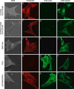
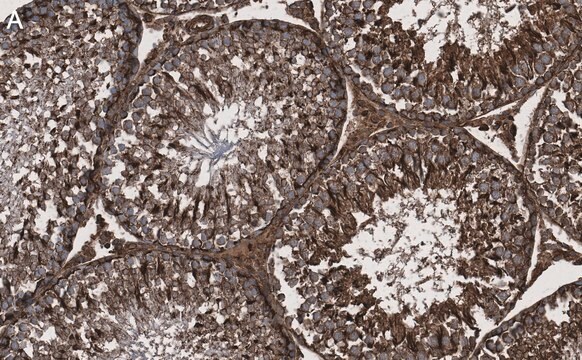
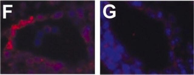
10μg/mL dilution from a previous lot was used for paraffin embedded, 4% formaldehyde, 3% glutaraldehyde, sodium cacodylate treated sections {see Luciano, L et al. 2003}.

ELISA:
A previous lot was shown to be strongly reactive with the cytoplasmic actin and shows a significant binding to gizzard, skeletal, arterial and cardiac actins. Also shows a significant binding to both Dictyostelium discoidum and Physarum polycephalum.

Western blot:
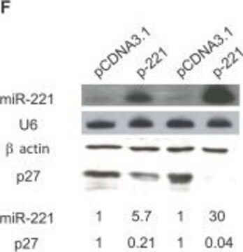
1-20 µg/ml. On muscle homogenates subject to SDS-PAGE, reacts relatively uniformly with a 43 kD protein present in skeletal, cardiac, gizzard and aorta tissues. Appears to react with all isoforms of actin found in these preparations and shows a strong reaction with the alpha-actin found in skeletal, cardiac, and arterial muscle (Otey, 1987).

Immunohistochemistry:
10µg/mL for paraffin embedded, 4% formaldehyde, 3% glutaraldehyde, sodium cacodylate treated sections {see Luciano, L et al. 2003}.

Optimal working dilutions must be determined by end user.
This Anti-Actin Antibody, clone C4 is validated for use in ELISA, IC, IH, IH(P), WB for the detection of Actin and has published in over 70 citations.

Quality

Routinely evaluated by Western Blot on HeLa lysate.

Western Blot Analysis:
1:1000 dilution of this lot detected actin on 10 μg of HeLa lysate.

Target description

43 kDa

Physical form

Protein G Purified in 0.1M Tris-Glycine (pH 7.4) 150mM NaCl with 0.05% NaN3.

Legal Information

CHEMICON is a registered trademark of Merck KGaA, Darmstadt, Germany

Not finding the right product?  

Try our Product Selector Tool.

Storage Class

12 - Non Combustible Liquids

wgk_germany

WGK 1

flash_point_f

Not applicable

flash_point_c

Not applicable


Certificates of Analysis (COA)

Search for Certificates of Analysis (COA) by entering the products Lot/Batch Number. Lot and Batch Numbers can be found on a product’s label following the words ‘Lot’ or ‘Batch’.

Already Own This Product?

Find documentation for the products that you have recently purchased in the Document Library.

Visit the Document Library

The ubiquitin-proteasome system is inhibited by p53 protein expression in human ovarian cancer cells.
Hwang IY, Baguley BC, Ching LM, Gilchrist CA
Cancer letters null
IgE-dependent sensitization increases responsiveness to LPS but does not modify development of endotoxin tolerance in mast cells.
Medina-Tamayo J, Ibarra-Sanchez A, Padilla-Trejo A, Gonzalez-Espinosa C.
Inflammation Research null
Increase in intracellular PGE2 induces apoptosis in Bax-expressing colon cancer cell.
Lalier, L; Pedelaborde, F; Braud, C; Menanteau, J; Vallette, FM; Olivier, C
BMC Cancer null
E Hervouet et al.
Cell death & disease, 1, e8-e8 (2010-01-01)
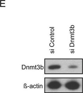
Disruption of apoptosis is considered as an important factor aiding tumorigenesis, and aberrant DNA methylation of apoptosis-associated genes could be an important and significant mechanism through which tumor cells avoid apoptosis. However, little is known about (1) the impact of
MMP-9 gene deletion mitigates microvascular loss in a model of ischemic acute kidney injury.
Lee, SY; Horbelt, M; Mang, HE; Knipe, NL; Bacallao, RL; Sado, Y; Sutton, TA
American Journal of Physiology: Renal Physiology null

Our team of scientists has experience in all areas of research including Life Science, Material Science, Chemical Synthesis, Chromatography, Analytical and many others.

Contact Technical Service