Skip to Content
MilliporeSigma
All Photos(1)

Key Documents

AB9610-AF555

Sigma-Aldrich

Anti-Olig-2 Antibody Alexa Fluor 555 Conjugate

from rabbit, ALEXA FLUOR 555

Synonym(s):

Oligodendrocyte transcription factor 2, bHLHe19, Olg-2, Oligo2, RACK17, RK17

Sign Into View Organizational & Contract Pricing


About This Item

UNSPSC Code:
12352203
eCl@ss:
32160702
NACRES:
NA.45

biological source

rabbit

Quality Level

conjugate

ALEXA FLUOR 555

antibody form

purified antibody

antibody product type

primary antibodies

clone

polyclonal

species reactivity

rat, mouse, human

technique(s)

immunofluorescence: suitable

NCBI accession no.

UniProt accession no.

shipped in

wet ice

target post-translational modification

unmodified

Gene Information

human ... OLIG2(10215)

General description

Oligodendrocyte transcription factor 2 (UniProt Q9EQW6; also known as bHLHe19, Olg-2, Oligo2, RACK17, RK17) is encoded by the Olig2 (also known as Bhlhb1) gene (Gene ID 50913) in murine species. Olig2 is a basic helix-loop-helix (bHLH) transcription factor expressed in a spinal cord ventricular zone domain where it mediates the sequential generation of motoneurons and oligodendrocytes during embryogenesis. Olig2 also functions as an anti-neurogenic factor in pMN progenitors at early time points to sustain the cycling progenitor pool. The anti-neurogenicity of Olig2 is also responsible for its role in tumorigenesis, especially in brain tumors and leukemia. Due to its expression in glioblastoma and other diffuse gliomas, Olig2 is a useful diagnostic marker of these brain tumors.

Specificity

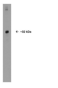
The unconjugated antibody (Cat. No. AB9610) recognizes the ~32 kDa Olig-2 protein by Western blotting.

Immunogen

Recombinant mouse Olig-2.

Application

Anti-Olig-2 Antibody Alexa Fluor 555 Conjugate is an antibody against Olig-2 for use in Immunofluorescence.
Research Category
Neuroscience
Research Sub Category
Transcription Factors
The unconjugated antibody (Cat. No. AB9610) is shown to be suitable also for Western blotting, immunocytochemistry, immunohistochemistry, and immunoprecipitation applications.

Quality

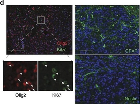
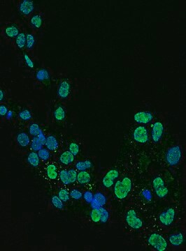
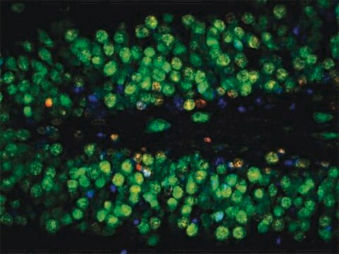
Evaluated by Immunofluorescence in mouse brain tissue.

Immunofluorescence Analysis: An 1:150 dilution of this antibody detected Olig-2 in mouse brain tissue.

Target description

~32 kDa observed.

Physical form

Ammonium Sulfate Precipitation
Purified rabbit polyclonal antibody conjugate in PBS with 15 mg/mL BSA and 0.1 % sodium azide.

Storage and Stability

Stable for 1 year at 2-8°C from date of receipt.

Other Notes

Concentration: Please refer to lot specific datasheet.

Legal Information

ALEXA FLUOR is a trademark of Life Technologies

Disclaimer

Unless otherwise stated in our catalog or other company documentation accompanying the product(s), our products are intended for research use only and are not to be used for any other purpose, which includes but is not limited to, unauthorized commercial uses, in vitro diagnostic uses, ex vivo or in vivo therapeutic uses or any type of consumption or application to humans or animals.

Not finding the right product?  

Try our Product Selector Tool.

Storage Class

12 - Non Combustible Liquids

wgk_germany

WGK 2

flash_point_f

Not applicable

flash_point_c

Not applicable


Certificates of Analysis (COA)

Search for Certificates of Analysis (COA) by entering the products Lot/Batch Number. Lot and Batch Numbers can be found on a product’s label following the words ‘Lot’ or ‘Batch’.

Already Own This Product?

Find documentation for the products that you have recently purchased in the Document Library.

Visit the Document Library

Isabel Mendizabal et al.
Genome biology, 20(1), 135-135 (2019-07-11)
The importance of cell type-specific epigenetic variation of non-coding regions in neuropsychiatric disorders is increasingly appreciated, yet data from disease brains are conspicuously lacking. We generate cell type-specific whole-genome methylomes (N = 95) and transcriptomes (N = 89) from neurons and oligodendrocytes obtained from
Ravian L van Ineveld et al.
Nature protocols, 17(12), 3028-3055 (2022-10-01)
Revealing the 3D composition of intact tissue specimens is essential for understanding cell and organ biology in health and disease. State-of-the-art 3D microscopy techniques aim to capture tissue volumes on an ever-increasing scale, while also retaining sufficient resolution for single-cell

Our team of scientists has experience in all areas of research including Life Science, Material Science, Chemical Synthesis, Chromatography, Analytical and many others.

Contact Technical Service