Skip to Content
MilliporeSigma
All Photos(3)

Key Documents

AB3848-I

Sigma-Aldrich

Anti-phospho Smad1/Smad5/Smad8 (Ser463/465)

from rabbit, purified by affinity chromatography

Synonym(s):

Mothers against decapentaplegic homolog 1, BSP-1, hSMAD1, JV4-1, MAD homolog 1, Mothers against DPP homolog 1, Mad-related protein 1, SMAD 1, SMAD family member 1, Smad1, Transforming growth factor-beta-signaling protein 1

Sign Into View Organizational & Contract Pricing


About This Item

UNSPSC Code:
12352203
eCl@ss:
32160702
NACRES:
NA.41

biological source

rabbit

Quality Level

antibody form

affinity isolated antibody

antibody product type

primary antibodies

clone

polyclonal

purified by

affinity chromatography

species reactivity

human, rat, mouse

species reactivity (predicted by homology)

Xenopus (based on 100% sequence homology), mink (based on 100% sequence homology)

technique(s)

immunohistochemistry: suitable (paraffin)
western blot: suitable

NCBI accession no.

UniProt accession no.

shipped in

ambient

target post-translational modification

phosphorylation (pSer463/pSer465)

Gene Information

human ... SMAD1(4086)

General description

Mothers against decapentaplegic homolog 1 (UniProt Q15797; also known as BSP-1, hSMAD1, JV4-1, MAD homolog 1, Mothers against DPP homolog 1, Mad-related protein 1, SMAD 1, SMAD family member 1, Smad1, Transforming growth factor-beta-signaling protein 1) is encoded by the SMAD1 (also known as BSP1, MADH1, MADR1) gene (Gene ID 4086) in human. Mammalian SMADs constitute a family of transcription factors that are subdivided into three groups. SMAD1, 2, 3, 5, and 9 (a.k.a. SMAD8) are receptor-regulated SMADs or R-SMADs. SMAD4 is the only known mammalian common mediator SMAD or Co-SMAD and it mediates both the TGF- and BMP signalling pathways. SMAD6 and SMAD7 are inhibitory SMADs or I-SMADs that block the activation of R-SMADs by Co-SMAD via competitive binding. SMADs share a similar structure feature consisting of the conserved N-terminal Mad-homology 1 (MH1; a.a. 12-136 of hSMAD1, a.a. 13-137 of hSMAD2, a.a. 16-140 of hSMAD8) and C-terminal MH2 (a.a. 271-465 of hSMAD1 & hSMAD2, a.a. 273-467 of hSMAD8) domains joined by a non-conserved linker region among SMADs. MH1 domain in R-SMADs and Co-SMADs contains N-terminus nuclear localization signals (NLS), while SMAD4 has nuclear export signals (NES) in this domain. The MH2 domain is responsible for gene transactivation as well as for mediating SMAD-receptor, SMAD-SMAD, SMAD-coactivators and SMAD-corepressors interactions. A TGF- ligand (TGF- for SMAD2/3 and BMP for SMAD1/5/8) initiates signalling by binding and engaging type I and type II receptor serine/threonine kinases on the cell surface, allowing T RII-dependent phosphorylation of T RI kinase domain, which then instigates signalling by phosphorylating the last two serine residues within the conserved C-terminal end Ser-Ser-X-Ser (SSXS) motif of SMAD proteins.

Specificity

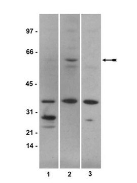
This rabbit polyclonal antibody recognizes SMAD1/5/8 phosphorylated on their last two serine residues within the C-terminal end Ser-Ser-X-Ser (SSXS) motif. Target serine residues are present in human/mouse SMAD1 (S463/S465), rat SMAD1 (S466/S468), human/mouse/rat SMAD5 (S463/S465), human SMAD9 (S465/S467), mouse SMAD9 (S428/S430), and rat SMAD9 (S432/S434), but absent in human SMAD1 spliced isoform 2. SMAD9 is also known as SMAD8. Target bands detection was blocked by the immunogen phosphopeptide, but not by the corresponding non-phosphopeptide.

Immunogen

Epitope: C-terminus
KLH-conjugated linear peptide corresponding to human SMAD1 C-terminal end sequence phosphorylated on the last two serine residues (S463/S465).

Application

Anti-phospho Smad1/Smad5/Smad8 (Ser463/465) Antibody, Cat. No. AB3848-I, is a highly specific rabbit polyclonal antibody that targets Smad1//5/8 phosphorylation and has been tested in Immunohistochemistry and Western Blotting.
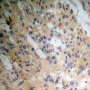
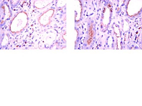
Immunohistochemistry Analysis: A 1:50 dilution from a representative lot detected Smad1/5/8 phosphorylation in mouse & rat embryo tissue sections.
Research Category
Epigenetics & Nuclear Function

Quality

Evaluated by Western Blotting in HEK293 cell lysates.

Western Blotting Analysis: A 1:500 dilution of this antibody detected an increased SMAD1/5/8 phosphorylation in 10 µg lysate from BMP2-treated HEK293 cells. Target bands detection was blocked by the immunogen phosphopeptide, but not the corresponding non-phosphopeptide.

Target description

~55 kDa observed. 52.26/52.16/52.71 kDa (human/mouse/rat SMAD1), 52.26/52.17/52.22 kDa (human/mouse/rat SMAD5), and 52.49/48.64/48.99 kDa Uncharacterized bands may be observed in some lysate(s).

Physical form

Affinity purified.
Purified rabbit polyclonal antibdoy in buffer containing 0.1 M Tris-Glycine (pH 7.4) 150 mM NaCl with 0.05% sodium azide

Storage and Stability

Stable for 1 year at 2-8°C from date of receipt.

Other Notes

Concentration: Please refer to lot specific datasheet.

Disclaimer

Unless otherwise stated in our catalog or other company documentation accompanying the product(s), our products are intended for research use only and are not to be used for any other purpose, which includes but is not limited to, unauthorized commercial uses, in vitro diagnostic uses, ex vivo or in vivo therapeutic uses or any type of consumption or application to humans or animals.

Not finding the right product?  

Try our Product Selector Tool.

Storage Class

12 - Non Combustible Liquids

wgk_germany

WGK 1

flash_point_f

Not applicable

flash_point_c

Not applicable


Certificates of Analysis (COA)

Search for Certificates of Analysis (COA) by entering the products Lot/Batch Number. Lot and Batch Numbers can be found on a product’s label following the words ‘Lot’ or ‘Batch’.

Already Own This Product?

Find documentation for the products that you have recently purchased in the Document Library.

Visit the Document Library

Yadong Liu et al.
Molecular medicine reports, 19(1), 461-467 (2018-11-30)
Lonicera japonica has been used in traditional Chinese medicine as an important medicinal plant, with the ability to inhibit osteoclast development and bone loss. However, it is not clear which active ingredient exerts these effects. (R)‑dehydroxyabscisic alcohol β‑D‑apiofuranosyl‑(1ˮ→6')‑β‑D‑glucopyranoside (DAG) is an
Xuguang Nie et al.
PLoS genetics, 14(7), e1007491-e1007491 (2018-07-06)
mTOR is a highly conserved serine/threonine protein kinase that is critical for diverse cellular processes in both developmental and physiological settings. mTOR interacts with a set of molecules including Raptor and Rictor to form two distinct functional complexes, namely the
Hongxiu Wen et al.
Biochimie, 160, 113-121 (2019-03-05)
Increased DAN protein (Grem1, Grem2, Grem3, Cerberus, NBL1, SOST, and USAG1) levels are often associated with severe disease-states in adult kidneys. Grem1, SOST, and USAG1 have been demonstrated to be upregulated and play a critical role in the progression of
Xiaohe Yan et al.
Investigative ophthalmology & visual science, 61(2), 44-44 (2020-02-28)
The clinical phenotype of retinal gliosis occurs in different forms; here, we characterize one novel genetic feature, (i.e., signaling via BMP-receptor 1b). Mouse mutants were generated within a recessive ENU mutagenesis screen; the underlying mutation was identified by linkage analysis
Marja M Hurley et al.
Scientific reports, 12(1), 15968-15968 (2022-09-25)
We have used Basic Fibroblast Growth Factor (FGF2) transgenic mice as experimental models for human X-linked hypophosphatemia (XLH)-related degenerative osteoarthritis (OA) to investigate the pathogenesis of the disease and to test potential pharmacotherapies for treatment. This study tested the efficacy

Our team of scientists has experience in all areas of research including Life Science, Material Science, Chemical Synthesis, Chromatography, Analytical and many others.

Contact Technical Service