跳轉至內容
Merck
全部照片(4)

Key Documents

P6556

Sigma-Aldrich

蛋白酶 K 来源于林伯氏白色念球菌

lyophilized powder, ≥30 units/mg protein

同義詞:

肽链内切酶

登入查看組織和合約定價


About This Item

CAS號碼:
酶委員會編號:
EC號碼:
MDL號碼:
分類程式碼代碼:
12352204
eCl@ss:
32160410
NACRES:
NA.54

形狀

lyophilized powder

品質等級

比活性

≥30 units/mg protein

分子量

28.93 kDa

技術

DNA extraction: suitable

溶解度

H2O: soluble 1 mg/mL, clear, colorless

異物活動

Dnase ≤30 Kunitz units/mg solid
RNase ≤0.003 Kunitz units/mg solid

運輸包裝

wet ice

儲存溫度

−20°C

尋找類似的產品? 前往 產品比較指南

應用

P6556产品为冻干粉形式。P6556产品已可用于分解人晶状体蛋白。蛋白酶K消化的蛋白酶印迹可以揭示蛋白质与蛋白质表面的相互作用。Sigma的该酶已用于鸡胚的预杂交。它还被用来富集PrPSc,一种存在于羊、仓鼠和老鼠痒病样本中的朊病毒蛋白。
在分离DNA和RNA的过程中,用于核酸酶的蛋白水解失活。
用于去除内毒素结合的阳离子蛋白,如溶菌酶和核糖核酸酶A。
据报道,其可用于分离肝、酵母和绿豆线粒体
用于测定酶在细胞膜上的定位
用于石蜡包埋组织切片的处理,暴露用于抗体标记的抗原结合位点。
用于可传播海绵状脑病(TSE)研究中朊病毒脑组织样本蛋白的消化。
在分离DNA和RNA的过程中,蛋白酶K对核酸酶的蛋白水解失活起作用。
它被用于去除内毒素结合的阳离子蛋白,如溶菌酶和核糖核酸酶A。
它可用于分离肝、酵母和绿豆线粒体
,并用于测定酶在细胞膜上的定位
用于石蜡包埋组织切片的处理,暴露用于抗体标记的抗原结合位点,并
用于可传播海绵状脑病(TSE)研究中朊病毒脑组织样本蛋白的消化。 P6556产品为冻干粉形式。 P6556产品已可用于分解人晶状体蛋白

生化/生理作用

蛋白酶K具有广泛的特异性,即使在天然状态下也能降解许多蛋白质。它主要裂解脂肪族和芳香族氨基酸羧基附近的含有封闭α-氨基的肽键。最佳pH值在7.5-9.0之间,等电点为8.9,其活性激活需要 Ca2+ (1-5 mM)。蛋白酶K可被二异丙基氟磷酸(DFIP)和苯甲磺酰氟(PMSF)抑制。
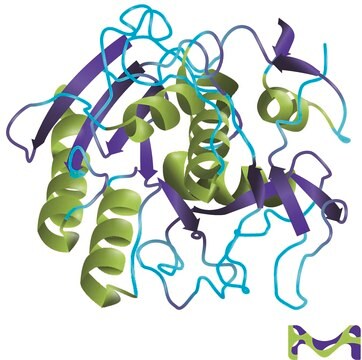
蛋白酶K是一种稳定且具有高活性的丝氨酸蛋白酶。晶体和分子结构研究证据表明该酶属于枯草素家族,具有活性位点催化三联体(Asp39-His69-Ser224)。它在一系列广泛的环境中均稳定:pH,缓冲盐,去污剂(SDS)以及温度。 在0.1-0.5% SDS存在的情况下,蛋白酶K可保持活性,在不损害分离DNA完整性的前提下,可以消化DNA制剂中的多种蛋白质和核酸酶。

單位定義

一单位在37°C、pH 7.5条件下每分钟可水解尿素变性血红蛋白而产生相当于1.0μmole(181 μg)的酪氨酸显色(福林酚试剂显色)。

也與該產品經常一起購買

產品號碼
描述
訂價

象形圖

Health hazardExclamation mark

訊號詞

Danger

危險分類

Eye Irrit. 2 - Resp. Sens. 1 - Skin Irrit. 2 - STOT SE 3

標靶器官

Respiratory system

儲存類別代碼

11 - Combustible Solids

水污染物質分類(WGK)

WGK 1

閃點(°F)

Not applicable

閃點(°C)

Not applicable

個人防護裝備

dust mask type N95 (US), Eyeshields, Faceshields, Gloves


分析證明 (COA)

輸入產品批次/批號來搜索 分析證明 (COA)。在產品’s標籤上找到批次和批號,寫有 ‘Lot’或‘Batch’.。

已經擁有該產品?

您可以在文件庫中找到最近購買的產品相關文件。

存取文件庫

M M Kristjánsson et al.
European journal of biochemistry, 260(3), 752-760 (1999-04-02)
An extracellular serine proteinase purified from cultures of a psychrotrophic Vibrio species (strain PA-44) belongs to the proteinase K family of the superfamily of subtilisin-like proteinases. The enzyme is secreted as a 47-kDa protein, but under mild heat treatment (30
Rongzhu Cheng et al.
The Journal of biological chemistry, 279(44), 45441-45449 (2004-08-19)
We report here the isolation of a novel acid-labile yellow chromophore from the enzymatic digest of human lens proteins and the identification of its chemical structure by liquid chromatography-mass spectrometry, liquid chromatography-tandem mass spectrometry, and (1)H, (13)C, and two-dimensional NMR.
Nathaniel Denkers et al.
Developmental dynamics : an official publication of the American Association of Anatomists, 229(3), 651-657 (2004-03-03)
Multi-color whole-mount in situ hybridization is a powerful technique for comparing the spatial expression patterns of two or more genes in developing embryos. We have developed an amplified triple-label whole-mount fluorescence in situ hybridization (FISH) protocol that permits detection of
Michela Candelma et al.
Reproduction (Cambridge, England), 153(2), 123-132 (2016-11-03)
In vertebrates, the regulation of gametogenesis is under the control of gonadotropins (Gth), follicle-stimulating hormone (Fsh) and luteinizing hormone (Lh). In fish, the physiological role of Gths is not fully understood, especially in species with asynchronous ovarian development. To elucidate
Janus S Jakobsen et al.
Science advances, 5(7), eaaw4304-eaaw4304 (2019-07-17)
The key myeloid transcription factor (TF), CEBPA, is frequently mutated in acute myeloid leukemia (AML), but the direct molecular effects of this leukemic driver mutation remain elusive. To investigate CEBPA mutant AML, we performed microscale, in vivo chromatin immunoprecipitation sequencing

文章

Proteinase K (EC 3.4.21.64) activity can be measured spectrophotometrically using hemoglobin as the substrate. Proteinase K hydrolyzes hemoglobin denatured with urea, and liberates Folin-postive amino acids and peptides. One unit will hydrolyze hemoglobin to produce color equivalent to 1.0 μmol of tyrosine per minute at pH 7.5 at 37 °C (color by Folin & Ciocalteu's Phenol Reagent).

條款

Proteinase K (EC 3.4.21.64) activity can be measured spectrophotometrically using hemoglobin as the substrate. Proteinase K hydrolyzes hemoglobin denatured with urea, and liberates Folin-postive amino acids and peptides. One unit will hydrolyze hemoglobin to produce color equivalent to 1.0 μmol of tyrosine per minute at pH 7.5 at 37 °C (color by Folin & Ciocalteu's Phenol Reagent).

In Situ Hybridization of Whole-Mount Mouse Embryos with RNA Probes: Hybridization, Washes, and Histochemistry

我們的科學家團隊在所有研究領域都有豐富的經驗,包括生命科學、材料科學、化學合成、色譜、分析等.

聯絡技術服務