Recommended Products
usage
sufficient for 100 tests
Quality Level
manufacturer/tradename
Calbiochem®
storage condition
OK to freeze
avoid repeated freeze/thaw cycles
technique(s)
immunoblotting: suitable
input
sample type cell extract(s)
storage temp.
−20°C
General description
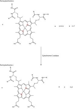
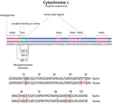
Assay kit provides a unique combination of reagents useful for the isolation of a highly enriched mitochondrial fraction from the cytosol. Translocation of cytochrome c from the mitochondrial fraction to the cytosol is monitored by immunoblotting with the cytochrome c antibody provided with the kit.
Components
Mitochondria Extraction Buffer, Cytosol Extraction Buffer, Protease Inhibitor Cocktail, Reducing Reagent, Cytochrome c Antibody, and a user protocol.
Warning
Toxicity: Multiple Toxicity Values, refer to MSDS (O)
Specifications
Assay Time: 4 h
Principle
The Cytochrome c Release Apoptosis Assay Kit provides a unique combination of reagents useful for the isolation of a highly enriched mitochondrial fraction from the cytosol that can be used to detect cytochrome c by Western blotting.
Preparation Note
• 1X Cytosol Extraction Buffer: Prepare 1X Cytosol Extraction Buffer by adding the 20 ml 5X Cytosol Extraction Buffer to 80 ml diH2O; mix well.• Mitochondria Extraction Buffer Mix: Just prior to performing the assay, prepare enough Mitochondria Extraction Buffer Mix for the number of samples to be assayed; each sample requires 0.1 ml of the mix. To prepare 1 ml Mitochondria Extraction Buffer Mix, add 2 µl 500X Protease Inhibitor Cocktail and 1 µl DTT to 1 ml Mitochondria Extraction Buffer; mix well.• Cytosol Extraction Buffer Mix: Just prior to performing the assay, prepare enough Cytosol Buffer Mix for the number of samples to be assayed; each sample requires 1 ml of the mix. To prepare 1 ml Cytosol Extraction Buffer Mix, add 2 µl 500X Protease Inhibitor Cocktail and 1 µl DTT to 1 ml Cytosol Extraction Buffer; mix well.
Storage and Stability
Upon arrival store the entire contents of the kit at -20°C. Once opened, store the Mitochondria Extraction Buffer and Cytosol Extraction Buffer at 4°C and the remaining components at -20°C.
Other Notes
Due to the nature of the Hazardous Materials in this shipment, additional shipping charges may be applied to your order. Certain sizes may be exempt from the additional hazardous materials shipping charges. Please contact your local sales office for more information regarding these charges.
Legal Information
CALBIOCHEM is a registered trademark of Merck KGaA, Darmstadt, Germany
Storage Class
8A - Combustible corrosive hazardous materials
Certificates of Analysis (COA)
Search for Certificates of Analysis (COA) by entering the products Lot/Batch Number. Lot and Batch Numbers can be found on a product’s label following the words ‘Lot’ or ‘Batch’.
Already Own This Product?
Find documentation for the products that you have recently purchased in the Document Library.
Syed Suhail Andrabi et al.
Disease models & mechanisms, 10(6), 787-796 (2017-04-02)
Organelle damage and increases in mitochondrial permeabilization are key events in the development of cerebral ischemic tissue injury because they cause both modifications in ATP turnover and cellular apoptosis/necrosis. Early restoration of blood flow and improvement of mitochondrial function might
Yin Liu et al.
Virology, 360(2), 364-375 (2006-12-13)
We previously showed that infection of rat oligodendrocytes by ultraviolet light-inactivated mouse hepatitis virus (MHV) resulted in apoptosis, suggesting that the apoptosis is triggered during cell entry. To further characterize the earliest apoptotic signaling events, here we treated cells with
Role of the mitochondrial signaling pathway in murine coronavirus-induced oligodendrocyte apoptosis.
Yin Liu et al.
Journal of virology, 80(1), 395-403 (2005-12-15)
A previous study demonstrated that infection of rat oligodendrocytes by mouse hepatitis virus (MHV) resulted in apoptosis, which is caspase dependent (Y. Liu, Y. Cai, and X. Zhang, J. Virol. 77:11952-11963, 2003). Here we determined the involvement of the mitochondrial
Gurdeep Marwarha et al.
Biomedicines, 10(1) (2022-01-22)
Apoptotic cell death of cardiomyocytes is a characteristic hallmark of ischemia-reperfusion (I/R) injury. The master hypoxamiR, microRNA-210 (miR-210), is considered the primary driver of the cellular response to hypoxic stress. However, to date, no consensus has emerged with regards to
Our team of scientists has experience in all areas of research including Life Science, Material Science, Chemical Synthesis, Chromatography, Analytical and many others.
Contact Technical Service


