16-156
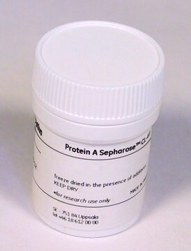
Protein A Agarose, Fast Flow
Protein A Agarose, Fast Flow suitable for medium and low-pressure chromatography, immunoprecipitation and antibody purification.
Synonym(s):
Protein A resin
Sign Into View Organizational & Contract Pricing
All Photos(1)
About This Item
UNSPSC Code:
41116133
eCl@ss:
32160801
NACRES:
NA.56
Recommended Products
form
liquid
manufacturer/tradename
Upstate®
technique(s)
affinity chromatography: suitable
immunoprecipitation (IP): suitable
western blot: suitable
shipped in
wet ice
General description
Protein A is an immunoglobulin (Ig)-binding protein used to purify large amounts of IgG. It binds to the Fc part of the antibody at the CH2–CH3 interface. Protein A-agarose might be suitable for low-pressure antibody isolation.
Recombinant Protein A covalently coupled to highly cross-linked 6% agarose beads.
Binding capacity: 40mg human IgG/ml agarose
Recombinant Protein A covalently coupled to highly cross-linked 6% agarose beads.
Binding capacity: 40mg human IgG/ml agarose
Application
Protein A Agarose, Fast Flow has been used in immunoprecipitation and chromatin immunoprecipitation (ChIP).
Quality
routinely evaluated in immunoprecipitation
Physical form
sterile distilled water containing 0.01% thimerosal
Legal Information
UPSTATE is a registered trademark of Merck KGaA, Darmstadt, Germany
Storage Class
10 - Combustible liquids
wgk_germany
WGK 1
Certificates of Analysis (COA)
Search for Certificates of Analysis (COA) by entering the products Lot/Batch Number. Lot and Batch Numbers can be found on a product’s label following the words ‘Lot’ or ‘Batch’.
Already Own This Product?
Find documentation for the products that you have recently purchased in the Document Library.
Customers Also Viewed
Simon Schenk et al.
American journal of physiology. Endocrinology and metabolism, 291(2), E254-E260 (2006-05-04)
Although the increase in fatty acid oxidation after endurance exercise training has been linked with improvements in insulin sensitivity and overall metabolic health, the mechanisms responsible for increasing fatty acid oxidation after exercise training are not completely understood. The primary
Ghia M Euskirchen et al.
Genome research, 17(6), 898-909 (2007-06-15)
Recent progress in mapping transcription factor (TF) binding regions can largely be credited to chromatin immunoprecipitation (ChIP) technologies. We compared strategies for mapping TF binding regions in mammalian cells using two different ChIP schemes: ChIP with DNA microarray analysis (ChIP-chip)
Lei Jiang et al.
Investigative ophthalmology & visual science, 61(5), 41-41 (2020-05-24)
To identify the pathogenic gene of infantile nystagmus syndrome (INS) in three Chinese families and explore the potential pathogenic mechanism of FERM domain-containing 7 (FRMD7) mutations. Genetic testing was performed via Sanger sequencing. Western blotting was used to analyze protein
Yong Cheng et al.
Nature, 515(7527), 371-375 (2014-11-21)
To broaden our understanding of the evolution of gene regulation mechanisms, we generated occupancy profiles for 34 orthologous transcription factors (TFs) in human-mouse erythroid progenitor, lymphoblast and embryonic stem-cell lines. By combining the genome-wide transcription factor occupancy repertoires, associated epigenetic
Vasavi Sundaram et al.
Genome research, 24(12), 1963-1976 (2014-10-17)
Transposable elements (TEs) have been shown to contain functional binding sites for certain transcription factors (TFs). However, the extent to which TEs contribute to the evolution of TF binding sites is not well known. We comprehensively mapped binding sites for
Our team of scientists has experience in all areas of research including Life Science, Material Science, Chemical Synthesis, Chromatography, Analytical and many others.
Contact Technical Service


