MABS1249
Przeciwciało anty-SCAP, klon 9D5
clone 9D5, from mouse
Synonim(y):
Sterol regulatory element-binding protein cleavage-activating protein, SCAP, SREBP cleavage-activating protein
About This Item
Polecane produkty
pochodzenie biologiczne
mouse
Poziom jakości
forma przeciwciała
purified immunoglobulin
rodzaj przeciwciała
primary antibodies
klon
9D5, monoclonal
reaktywność gatunkowa
hamster
metody
immunocytochemistry: suitable
western blot: suitable
izotyp
IgG2bκ
numer dostępu NCBI
numer dostępu UniProt
docelowa modyfikacja potranslacyjna
unmodified
informacje o genach
hamster ... Scap(100689048)
Opis ogólny
4 regionami lumenalnymi i 5 cytoplazmatycznymi, mając zarówno N-, jak i C-końcowe końce po stronie cytoplazmatycznej (a.a. 1-18 i 730-1276). C-końcowa domena SCAP pośredniczy w wiązaniu z SREBP, podczas gdy transbłonowe helisy 2-6 obejmują domenę wyczuwającą sterol, która pośredniczy w indukowanym sterolem wiązaniu SCAP z Insigs. Mutacje w regionie wyczuwającym sterol zakłócają wiązanie Insig i zapobiegają retencji SCAP-SREBP w ER za pośrednictwem sterolu.
Specyficzność
Immunogen
Zastosowanie
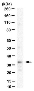
Analiza Western Blotting: Reprezentatywna partia wykryła egzogennie wyrażony SCAP koimmunoprecypitowany z egzogennie wyrażonym Insig-1 i Insig-2 po traktowaniu 25-hydroksycholesterolem (25-HC) linii komórkowej CHO z niedoborem SCAP SRD-13A, która została transfekowana w celu koekspresji SCAP z Insig-1 lub Insig-2 znakowanym myc (Lee, P.C. i DeBose-Boyd, R.A. (2010). J. Lipid Res. 51(1):192-201).
Analiza Western Blotting: Reprezentatywne partie wykryły podobny poziom SCAP w ekstraktach błonowych z komórek CHO z lub bez leczenia inhibitorem przetwarzania SREBP 25-hydroksycholesterolem (25-HC), niezależnie od tego, czy komórki są odporne na 25-HC (Lee, P.C. i DeBose-Boyd, R.A. (2010). J. Lipid Res. 51(1):192-201; Lee, P.C., et al. (2007). J. Lipid Res. 48(9):1944-1954).
Analiza Western Blotting: Reprezentatywna partia wykryła endogenny SCAP immunoprecypitowany z lizatu komórek CHO przez poliklonalne przeciwciało SCAP (Sakai, J., et al. (1997). J. Biol. Chem. 272(32):20213-20221).
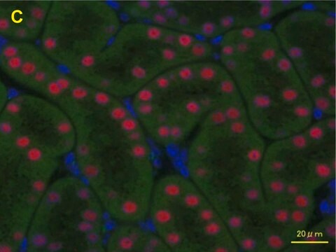
Analiza immunocytochemiczna: Reprezentatywna partia zlokalizowała nadeksprymowany SCAP chomika w ER i otoczce jądrowej przez fluorescencyjne barwienie immunocytochemiczne 3% utrwalonych paraformaldehydem, 0.01% przepuszczalnych dla saponiny stabilnych transfekantów CHO (Sakai, J., et al. (1997). J. Biol. Chem. 272(32):20213-20221).
Jakość
Isotyping Analysis: The identity of this monoclonal antibody is confirmed by isotyping test to be IgG2bκ.
Opis wartości docelowych
Postać fizyczna
Inne uwagi
Nie możesz znaleźć właściwego produktu?
Wypróbuj nasz Narzędzie selektora produktów.
Kod klasy składowania
12 - Non Combustible Liquids
Klasa zagrożenia wodnego (WGK)
WGK 1
Temperatura zapłonu (°F)
Not applicable
Temperatura zapłonu (°C)
Not applicable
Certyfikaty analizy (CoA)
Poszukaj Certyfikaty analizy (CoA), wpisując numer partii/serii produktów. Numery serii i partii można znaleźć na etykiecie produktu po słowach „seria” lub „partia”.
Masz już ten produkt?
Dokumenty związane z niedawno zakupionymi produktami zostały zamieszczone w Bibliotece dokumentów.
Nasz zespół naukowców ma doświadczenie we wszystkich obszarach badań, w tym w naukach przyrodniczych, materiałoznawstwie, syntezie chemicznej, chromatografii, analityce i wielu innych dziedzinach.
Skontaktuj się z zespołem ds. pomocy technicznej