Przejdź do zawartości
Merck
Wszystkie zdjęcia(1)

Key Documents

MABF939

Sigma-Aldrich

Anti-IFNAR1 Antibody, clone 4G8

clone 4G8, from mouse

Synonim(y):

Interferon alpha/beta receptor 1, CRF2-1, Cytokine receptor class-II member 1, Cytokine receptor family 2 member 1, IFN-alpha/beta receptor 1, IFN-R-1, Type I interferon receptor 1

Zaloguj sięWyświetlanie cen organizacyjnych i kontraktowych


About This Item

Kod UNSPSC:
12352203
eCl@ss:
32160702

pochodzenie biologiczne

mouse

Poziom jakości

forma przeciwciała

purified immunoglobulin

rodzaj przeciwciała

primary antibodies

klon

4G8, monoclonal

reaktywność gatunkowa

human

metody

flow cytometry: suitable

izotyp

IgG1κ

numer dostępu NCBI

numer dostępu UniProt

Warunki transportu

ambient

docelowa modyfikacja potranslacyjna

unmodified

informacje o genach

human ... IFNAR1(3454)

Opis ogólny

Interferon alpha/beta receptor 1 (UniProt P17181; also known as CRF2-1, Cytokine receptor class-II member 1, Cytokine receptor family 2 member 1, IFN-alpha/beta receptor 1, IFN-R-1, Type I interferon receptor 1) is encoded by the IFNAR1 (also known as IFNAR) gene (Gene ID 3454) in human. Interferons (IFNs) are cytokines whose prouction and release by host cells are induced in the presence of pathogens (e.g. viruses, bacteria, parasites) and tumor cells. More than twenty distinct IFN genes and proteins have been identified and classified into three main types. Type I IFNs (IFN-α, IFN-β, IFN-ε, IFN-κ, and IFN-ω) exert their function via binding IFN-α/β receptor (encoded by the IFNAR1 and IFNAR2 gene, respectively). Ligation of IFNAR by IFN-I results in activation of the Janus kinases, Jak1 and Tyk2, which in turn phosphorylates STAT1 and STAT2, resulting in recruitment of IRF9 to form the IFN-stimulated gene factor 3 (ISGF3) transcription factor. ISGF3 then translocates to the nucleus, where it binds IFN-stimulated response elements (ISREs) and drives the transcription of IFN-stimulated genes (ISGs). IFNAR1 is phosphorylated by Tyk2 and PKD2 upon IFN-I stimulation, leading to IFNAR1 ubiquitination and lysosomal degradation. IFNAR1 is also
phosphorylated by p38 MAP kinase in response to non-IFN stimuli, including the PERK-dependent unfolded protein response (UPR), ligation of pattern recognition receptors (PRRs), or through signaling via other inflammatory cytokines or growth factors including VEGF, IL-1β and TNFα. Encephalitic flaviviruses antagonize IFN-I signaling by inhibiting IFNAR1 surface expression, where the viral nonstructural protein 5 (NS5) targets cellular prolidase (PEPD) that is required for IFNAR1 maturation and accumulation.

Specyficzność

Clone 4G8 detected a loss of HEK293 cell surface IFNAR1 immunoreactivity following lentivirus-mediated cellular IFNAR1 shRNA delivery or West Nile virus (WNV) infection (Lubick, K.J., et al. (2015). Cell Host Microbe. 18(1):61-74).

Immunogen

Epitope: extracellular domain
His-tagged recombinant human IFNAR1 extracellular domain.

Zastosowanie

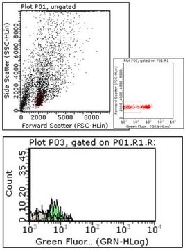
Flow Cytometry Analysis: A representative lot detected a time-dependent loss of HEK293 cell surface IFNAR1 immunoreactivity following West Nile virus (WNV) infection. A significantly faster surface IFNAR1 loss was observed when cells were infected with WNV that produces IFN-I-antagonizing NS5 (nonstructural protein 5) with S653F mutation (Lubick, K.J., et al. (2015). Cell Host Microbe. 18(1):61-74).

Flow Cytometry Analysis: A representative lot detected a loss of HEK293 cell surface IFNAR1 immunoreactivity following lentivirus-mediated cellular IFNAR1 shRNA delivery (Lubick, K.J., et al. (2015). Cell Host Microbe. 18(1):61-74).
Research Category
Inflammation & Immunology
This mouse monoclonal Anti-IFNAR1 Antibody, clone 4G8, Cat. No. MABF939 is validated for use in Flow Cytometry for the detection of IFNAR1.

Jakość

Evaluated by Flow Cytometry in K562 cells.

Flow Cytometry Analysis: 1 µg of this antibody detected IFNAR1 on the surface of K562 cells.

Opis wartości docelowych

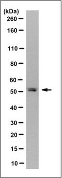
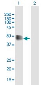
63.53/60.93 kDa (pro-/mature isoform 1), 49.55/46.95 kDa (pro-/mature isoform 2), 56.72/54.12 kDa (pro-/mature isoform 3), 56.05 kDa (isoform 4) calculated. Apparent molecular weight appears greater than 100 kDa due to glycosylation.

Postać fizyczna

Format: Purified
Protein G purified.
Purified mouse IgG1κ in buffer containing 0.1 M Tris-Glycine (pH 7.4), 150 mM NaCl with 0.05% sodium azide.

Przechowywanie i stabilność

Stable for 1 year at 2-8°C from date of receipt.

Inne uwagi

Concentration: Please refer to lot specific datasheet.

Oświadczenie o zrzeczeniu się odpowiedzialności

Unless otherwise stated in our catalog or other company documentation accompanying the product(s), our products are intended for research use only and are not to be used for any other purpose, which includes but is not limited to, unauthorized commercial uses, in vitro diagnostic uses, ex vivo or in vivo therapeutic uses or any type of consumption or application to humans or animals.
This page may contain text that has been machine translated.

Nie możesz znaleźć właściwego produktu?  

Wypróbuj nasz Narzędzie selektora produktów.

Kod klasy składowania

12 - Non Combustible Liquids

Klasa zagrożenia wodnego (WGK)

WGK 1

Temperatura zapłonu (°F)

Not applicable

Temperatura zapłonu (°C)

Not applicable


Certyfikaty analizy (CoA)

Poszukaj Certyfikaty analizy (CoA), wpisując numer partii/serii produktów. Numery serii i partii można znaleźć na etykiecie produktu po słowach „seria” lub „partia”.

Masz już ten produkt?

Dokumenty związane z niedawno zakupionymi produktami zostały zamieszczone w Bibliotece dokumentów.

Odwiedź Bibliotekę dokumentów

Nasz zespół naukowców ma doświadczenie we wszystkich obszarach badań, w tym w naukach przyrodniczych, materiałoznawstwie, syntezie chemicznej, chromatografii, analityce i wielu innych dziedzinach.

Skontaktuj się z zespołem ds. pomocy technicznej