Przejdź do zawartości
Merck
Wszystkie zdjęcia(2)

Kluczowe dokumenty

MABC44

Sigma-Aldrich

Anti-LAMP-3 Antibody, clone 16H11.2

clone 16H11.2, from mouse

Synonim(y):

LAMP, CD208, LAMP-3, DC-loysosome-associated membrane glycoprotein, DC LAMP, DC-LAMP, Protein TSC403

Zaloguj sięWyświetlanie cen organizacyjnych i kontraktowych


About This Item

Kod UNSPSC:
12352203
eCl@ss:
32160702
NACRES:
NA.41

pochodzenie biologiczne

mouse

Poziom jakości

forma przeciwciała

purified immunoglobulin

rodzaj przeciwciała

primary antibodies

klon

16H11.2, monoclonal

reaktywność gatunkowa

human

metody

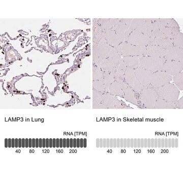
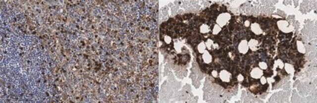
immunohistochemistry: suitable
western blot: suitable

izotyp

IgG2bκ

numer dostępu NCBI

numer dostępu UniProt

Warunki transportu

wet ice

docelowa modyfikacja potranslacyjna

unmodified

informacje o genach

human ... LAMP3(27074)

Opis ogólny

Lysosyme-associated membrane proteins (LAMP) are transmembrane lysosomal glycoproteins, which share a common Gly-Tyr motif as a lysosomal targeting signal. While LAMP-1 and LAMP-2 contain type 1 transmembrane domains, LAMP-3 contains a transmembrane 4 superfamily domain, tetraspanin. FceRI-mediated stimulation of basophils causes LAMP-3 to be translocated from its intracellular localization to the plasma membrane. LAMP-3 thereby serves as a reliable marker for basophil activation. LAMP-3 expression is known to be reported in human bone marrow mast cells and in leukemic human mast cell line (HMC-1), among other cell lines. LAMP3 is shown to be regulated by hypoxia in a panel of tumor cells, via the unfolded protein response (UPR). UPR is a mechanism of adaptation to endoplasmic reticulum stress and is demonstrated to contribute to hypoxic adaptation in tumors through multiple mechanisms.

Immunogen

Recombinant protein corresponding to human LAMP-3.

Zastosowanie

Detect LAMP-3 using this Anti-LAMP-3 Antibody, clone 16H11.2 validated for use in WB, IH.
Immunohistochemistry Analysis: A 1:200 dilution from a representative lot detected LAMP-3 in colorectal adenocarcinoma tissue.
Research Category
Cell Structure
Research Sub Category
Apoptosis - Additional

Organelle & Cell Markers

Jakość

Evaluated by Western Blot in MCF-7 cell lysate.

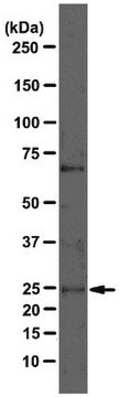
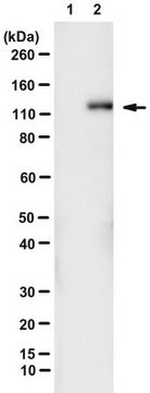
Western Blot Analysis: 1 µg/mL of this antibody detected LAMP-3 in MCF-7 cell lysate.

Opis wartości docelowych

~65 kDa observed.
The calculated molecular weight of this protein is 41 kDa, but has been observed at ~65-90 kDa due to glycosolation (de Saint-Vis, B., et al. (1998). Immunity. 9(3):325-336).

Postać fizyczna

Format: Purified
Protein G Purified
Purified mouse monoclonal IgG2bκ in buffer containing 0.1 M Tris-Glycine (pH 7.4), 150 mM NaCl with 0.05% sodium azide.

Przechowywanie i stabilność

Stable for 1 year at 2-8°C from date of receipt.

Komentarz do analizy

Control
MCF-7 cell lysate

Inne uwagi

Concentration: Please refer to the Certificate of Analysis for the lot-specific concentration.

Oświadczenie o zrzeczeniu się odpowiedzialności

Unless otherwise stated in our catalog or other company documentation accompanying the product(s), our products are intended for research use only and are not to be used for any other purpose, which includes but is not limited to, unauthorized commercial uses, in vitro diagnostic uses, ex vivo or in vivo therapeutic uses or any type of consumption or application to humans or animals.
Ta strona może zawierać tekst przetłumaczony maszynowo.

Nie możesz znaleźć właściwego produktu?  

Wypróbuj nasz Narzędzie selektora produktów.

Kod klasy składowania

12 - Non Combustible Liquids

Klasa zagrożenia wodnego (WGK)

WGK 1

Temperatura zapłonu (°F)

Not applicable

Temperatura zapłonu (°C)

Not applicable


Certyfikaty analizy (CoA)

Poszukaj Certyfikaty analizy (CoA), wpisując numer partii/serii produktów. Numery serii i partii można znaleźć na etykiecie produktu po słowach „seria” lub „partia”.

Masz już ten produkt?

Dokumenty związane z niedawno zakupionymi produktami zostały zamieszczone w Bibliotece dokumentów.

Odwiedź Bibliotekę dokumentów

Junya Nishimura et al.
Esophagus : official journal of the Japan Esophageal Society, 16(4), 333-344 (2019-04-11)
Dendritic cells (DCs) are the most potent antigen-presenting cells to induce cytotoxic T lymphocytes in the tumor environment. After acquiring antigens, DCs undergo maturation and their expression of MHC and co-stimulation molecules are enhanced, along with lysosome-associated membrane glycoprotein 3

Nasz zespół naukowców ma doświadczenie we wszystkich obszarach badań, w tym w naukach przyrodniczych, materiałoznawstwie, syntezie chemicznej, chromatografii, analityce i wielu innych dziedzinach.

Skontaktuj się z zespołem ds. pomocy technicznej