
MAB3430
Anti-Desmin Antibody, clone DE-B-5
clone DE-B-5, Chemicon®, from mouse
Zaloguj sięWyświetlanie cen organizacyjnych i kontraktowych
About This Item
Kod UNSPSC:
12352203
eCl@ss:
32160702
NACRES:
NA.41
Polecane produkty


pochodzenie biologiczne
mouse
Poziom jakości
forma przeciwciała
purified antibody
klon
DE-B-5, monoclonal
reaktywność gatunkowa
mouse, frog, pig, rat, human
producent / nazwa handlowa
Chemicon®
metody
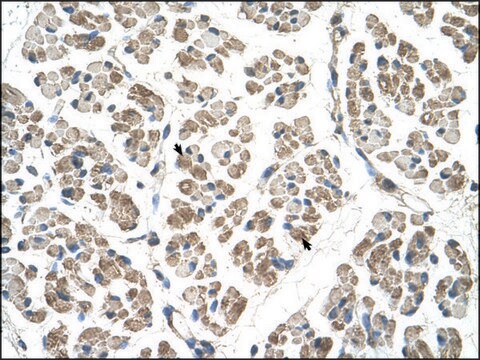
immunohistochemistry (formalin-fixed, paraffin-embedded sections): suitable
western blot: suitable
izotyp
IgG1
numer dostępu NCBI
numer dostępu UniProt
Warunki transportu
wet ice
docelowa modyfikacja potranslacyjna
unmodified
informacje o genach
human ... DES(1674)
Specyficzność
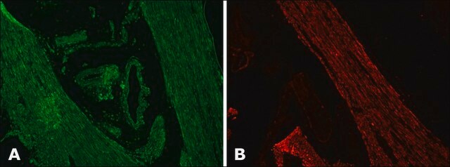
The antibody reacts with desmin from human, pig, rat and taod. In tissue sections this antibody is used to stain skeletal, cardiac, visceral, and some vascular smooth muscle cells. Cell lines such as RD (ATCC CCL 136) and hamster BHK-21 are positive (Debus et al., 1983).
Immunogen
Purified desmin.
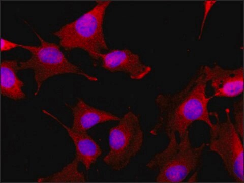
Zastosowanie
Detect Desmin using this Anti-Desmin Antibody, clone DE-B-5 validated for use in WB, IH, IH(P), IH(P).
Immunocytochemistry: (5 μg/ml) Immunohistochemistry: (5 μg/ml) See IHC2011-6 for prediluted and detailed paraffin protocols.
Optimal working dilutions must be determined by end user.
Immunohysto/cyto chemistry Protocols
Ideal specimens are obtained from frozen sections from shock-frozen tissue samples. The frozen sections are dried in the air and then fixed with acetone at -20°C for 10 min. Excess acetone is allowed to evaporate at 15-25°C. Material fixed in alcohol and embedded in paraffin can also be used (2). Formaldehyde fixation will reduce or eliminate the intensity of staining depending on the conditions under which it is performed. Other fixation conditions must be first tested by the investigator.
It is advantageous to block unspecific binding sites by overlaying the sections with fetal calf serum for 20-30 min at 15-25°C. Excess of fetal calf serum is removed by decanting before application of the antibody solution.
Cytocentrifuge preparations of single cells or cell smears are also fixed in acetone. These preparations should, however not be dried in the air. Instead, the excess acetone is removed by briefly washing in phosphate-buffered saline (PBS).
Further treatment is then as follows:
• Overlay the preparation with 10-20 μl antibody solution and incubate in a humid chamber at 37°C for 1 h.
• Dip the slide briefly in PBS and then wash 3 x in PBS for 3 min (using a fresh PBS bath in each case).
• Wipe the margins of the preparation dry and overlay the preparation with 10-20 μl of a solution of anti-mouse Ig-FITC or anti-mouse IgG-peroxidase solution and allow to incubate for 1 h at 37°C in a humid chamber.
• Wash the slide as described above.
The preparation must not be allowed to dry out during any of the steps.
If using an indirect immunofluorescence technique, the preparation should be overlaid with a suitable embedding medium (e.g. Moviol, Hoechst) and examined under the fluorescence microscope. If a POD-conjugate has been used as the secondary antibody, the preparation should be overlaid with a substrate solution (see below) and incubated at 15-25°C until a clearly visible redbrown color develops. A negative control (e.g. only the secondary antibody) should remain unchanged in color during this incubation period. Subsequently, the substrate is washed off with PBS and the preparation is stained, if desired, with hemalum stain for about 1 min. The hemalum solution is washed off with PBS; the preparation is embedded and examined.
Substrate solutions:
Aminoethyl-carbazole: Dissolve 2 mg 3-amino-9-ethylcarbazole with 1.2 ml dimethylsulfoxide and add 28.8 ml 50 mM Tris-HCI, pH 7.3, and 20 μl 3% H 2 O 2 (w/v). Prepare solution freshly each day. Diaminobenzidine: Dissolve 25 mg 3,3′-diaminobenzidine with 50 ml 50 mM Tris-HCI, pH 7.3, and add 40 μl 3% H 2 O 2 (w/v). Prepare solution freshly each day.
Research Category
Cell Structure
Cell Structure
Research Sub Category
Cytoskeleton
Cytoskeleton
Jakość
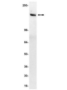
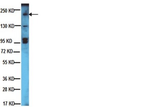
Western Blot Analysis: 1:1000 dilution
Powiązanie
Replaces: 04-585
Postać fizyczna
Format: Purified
Liquid in 0.02 M phosphate buffer, 0.25M NaCl with 0.1% sodium azide, pH 7.6.
Przechowywanie i stabilność
Maintain antibody refrigerated at 2-8°C in undiluted aliquots for up to 6 months. DO NOT FREEZE.
Inne uwagi
Concentration: Please refer to the Certificate of Analysis for the lot-specific concentration.
Informacje prawne
CHEMICON is a registered trademark of Merck KGaA, Darmstadt, Germany
Oświadczenie o zrzeczeniu się odpowiedzialności
Unless otherwise stated in our catalog or other company documentation accompanying the product(s), our products are intended for research use only and are not to be used for any other purpose, which includes but is not limited to, unauthorized commercial uses, in vitro diagnostic uses, ex vivo or in vivo therapeutic uses or any type of consumption or application to humans or animals.
Ta strona może zawierać tekst przetłumaczony maszynowo.
polecane
Kod klasy składowania
12 - Non Combustible Liquids
Klasa zagrożenia wodnego (WGK)
WGK 2
Temperatura zapłonu (°F)
Not applicable
Temperatura zapłonu (°C)
Not applicable
Certyfikaty analizy (CoA)
Poszukaj Certyfikaty analizy (CoA), wpisując numer partii/serii produktów. Numery serii i partii można znaleźć na etykiecie produktu po słowach „seria” lub „partia”.
Masz już ten produkt?
Dokumenty związane z niedawno zakupionymi produktami zostały zamieszczone w Bibliotece dokumentów.
Two-dimensional gel electrophoresis maps of the proteome and phosphoproteome of primitively cultured rat mesangial cells.
Xiao-Sheng Jiang,Liu-Ya Tang,Xing-Jun Cao,Hu Zhou,Qi-Chang Xia,Jia-Rui Wu,Rong Zeng
Electrophoresis null
Nathan R Tucker et al.
Experimental cell research, 315(18), 3176-3186 (2009-07-08)
Injury to muscle tissue plays a central role in various cardiovascular pathologies. Overexpression of the small heat shock protein Hsp27 protects muscle cells against thermal, oxidative and ischemic stress. However, underlying mechanisms of this protection have not been resolved. A
Wolfgang M Schmidt et al.
PLoS genetics, 7(4), e1002042-e1002042 (2011-05-03)
Albeit genetically highly heterogeneous, muscular dystrophies (MDs) share a convergent pathology leading to muscle wasting accompanied by proliferation of fibrous and fatty tissue, suggesting a common MD-pathomechanism. Here we show that mutations in muscular dystrophy genes (Dmd, Dysf, Capn3, Large)
Identification, selection, and enrichment of cardiomyocyte precursors.
Zanetti, BF; Gomes, WJ; Han, SW
BioMed Research International null
Fan Yang et al.
Journal of cellular physiology, 233(2), 1601-1611 (2017-06-22)
The estrogen-related receptor b (ESRRB) is an orphan nuclear receptor and targets many genes involved in self-renewal and pluripotency. In mouse ES cells, overexpression of ESRRB can maintain LIF-independent self-renewal in the absence of Nanog. However, the fundamental features of
Nasz zespół naukowców ma doświadczenie we wszystkich obszarach badań, w tym w naukach przyrodniczych, materiałoznawstwie, syntezie chemicznej, chromatografii, analityce i wielu innych dziedzinach.
Skontaktuj się z zespołem ds. pomocy technicznej