
추천 제품


생물학적 소스
rat
Quality Level
항체 형태
purified immunoglobulin
항체 생산 유형
primary antibodies
클론
TROMA-3, monoclonal
종 반응성
mouse, human
포장
antibody small pack of 25 μg
기술
electron microscopy: suitable
immunofluorescence: suitable
immunohistochemistry: suitable (paraffin)
immunoprecipitation (IP): suitable
western blot: suitable
동형
IgG2aκ
NCBI 수납 번호
UniProt 수납 번호
배송 상태
ambient
타겟 번역 후 변형
unmodified
유전자 정보
human ... KRT19(3880)
mouse ... Krt19(16669)
일반 설명
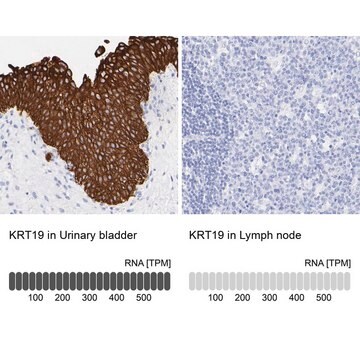
Keratin, type I cytoskeletal 19 (UniProt: P19001; also known as Cytokeratin-19, CK-19, Keratin-19, K19) is encoded by the Krt19 (also known as Krt1-19) gene (Gene ID: 16669) in murine species. CK-19 belongs to the family of intermediate filament (IF) family, but differs from other IF proteins in lacking the C-terminal tail domain. About 20 different cytokeratin proteins have been reported in higher animals in varying combinations during various stages of development. CK-19 is normally expressed in the lining of the gastroenteropancreatic and hepatobiliary tracts. It is expressed throughout embryonic development with highest levels observed at 8.5 days post coitum. Its expression is shown to decrease by day 11.5 and then is increased again by 17.5 days post coitum. CK-19 is shown to be involved in the organization of myofibrils and together with keratin 8, it helps to link the contractile apparatus to dystrophin at the costameres of striated muscle. CK-19 has been shown to be an independent prognostic factor for pancreatic neuroendocrine tumors, especially the insulin-negative tumors. TROMA 3 has been found to stain trophoblastoma TDM.1 cells, a subpopulation of the retinoic acid.-induced F9 and PCC3/ND1 cells, and in 5-azacytidine-treated 1246 cells.
특이성
Clone TROMA-3 detects cytokeratin 19 in human and murine cells.
면역원
Purified intermediate filaments from DM1 (Trophoblatoma) cell line.
애플리케이션
Anti-Cytokeratin 19, clone TROMA-3, Cat. No. MABT913, is a rat monoclonal antibody that detects Keratin, type I cytoskeletal 19 and is tested for use in Electron Microscopy, Immunofluorescence, Immunohistochemistry (Paraffin), Immunoprecipitation, and Western Blotting.
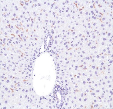
Immunohistochemistry Analysis: A 1:250 dilution from a representative lot detected Cytokeratin 19 in mouse lung tissue.
Western Blotting Analysis: A representative lot detected Cytokeratin 19 in Western Blotting applications (Boller, K., et. al. (1987). Eur J Cell Biol. 43(3):459-68).
Electron Microscopy Analysis: A representative lot detected Cytokeratin 19 in Electron Microscopy applications (Boller, K., et. al. (1987). Eur J Cell Biol. 43(3):459-68).
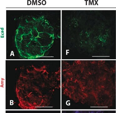
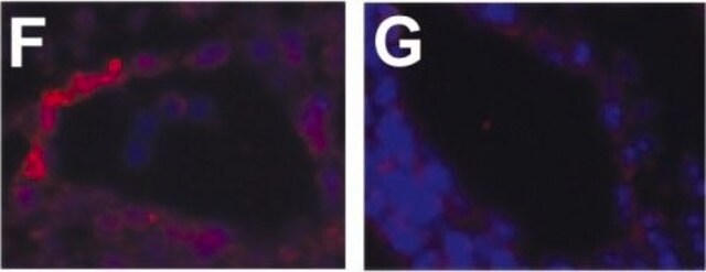
Immunofluorescence Analysis: A representative lot detected Cytokeratin 19 in Immunofluorescence applications (Boller, K., et. al. (1987). Eur J Cell Biol. 43(3):459-68).
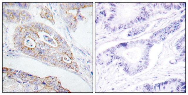
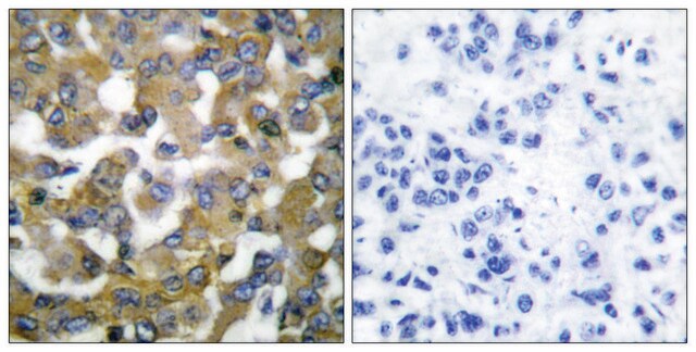
Immunohistochemistry Analysis: A representative lot detected Cytokeratin 19 in Immunohistochemistry applications (Takase, H.M., et. al. (2013). Genes Dev. 27(2):169-81).
Immunoprecipitation Analysis: A representative lot detected Cytokeratin 19 in Immunoprecipitation applications (Ichinose, Y., et. al. (1990). Biochem Biophys Res Commun. 167(2):644-7).
Western Blotting Analysis: A representative lot detected Cytokeratin 19 in Western Blotting applications (Boller, K., et. al. (1987). Eur J Cell Biol. 43(3):459-68).
Electron Microscopy Analysis: A representative lot detected Cytokeratin 19 in Electron Microscopy applications (Boller, K., et. al. (1987). Eur J Cell Biol. 43(3):459-68).
Immunofluorescence Analysis: A representative lot detected Cytokeratin 19 in Immunofluorescence applications (Boller, K., et. al. (1987). Eur J Cell Biol. 43(3):459-68).
Immunohistochemistry Analysis: A representative lot detected Cytokeratin 19 in Immunohistochemistry applications (Takase, H.M., et. al. (2013). Genes Dev. 27(2):169-81).
Immunoprecipitation Analysis: A representative lot detected Cytokeratin 19 in Immunoprecipitation applications (Ichinose, Y., et. al. (1990). Biochem Biophys Res Commun. 167(2):644-7).
품질
Evaluated by Western Blotting in MCF-7 cell lysate.
Western Blotting Analysis: 0.5 µg/mL of this antibody detected Cytokeratin 19 in 10 µg of MCF-7 cell lysate.
Western Blotting Analysis: 0.5 µg/mL of this antibody detected Cytokeratin 19 in 10 µg of MCF-7 cell lysate.
표적 설명
~44 kDa observed; 44.54 kDa calculated. Uncharacterized bands may be observed in some lysate(s).
물리적 형태
Format: Purified
기타 정보
Concentration: Please refer to lot specific datasheet.
적합한 제품을 찾을 수 없으신가요?
당사의 제품 선택기 도구.을(를) 시도해 보세요.
Storage Class Code
12 - Non Combustible Liquids
WGK
WGK 1
Flash Point (°F)
Not applicable
Flash Point (°C)
Not applicable
시험 성적서(COA)
제품의 로트/배치 번호를 입력하여 시험 성적서(COA)을 검색하십시오. 로트 및 배치 번호는 제품 라벨에 있는 ‘로트’ 또는 ‘배치’라는 용어 뒤에서 찾을 수 있습니다.
Ana Caroline Costa-da-Silva et al.
iScience, 25(1), 103592-103592 (2022-01-11)
Chronic graft-versus-host disease (cGVHD) targets include the oral mucosa and salivary glands after allogeneic hematopoietic stem cell transplant (HSCT). Without incisional biopsy, no diagnostic test exists to confirm oral cGVHD. Consequently, therapy is often withheld until severe manifestations develop. This
Rebecca Marcus et al.
Cancers, 13(22) (2021-11-28)
Intrahepatic cholangiocarcinoma (ICC) is a primary biliary malignancy that harbors a dismal prognosis. Oncogenic mutations of KRAS and loss-of-function mutations of BRCA1-associated protein 1 (BAP1) have been identified as recurrent somatic alterations in ICC. However, an autochthonous genetically engineered mouse
Romina Lasagni Vitar et al.
Stem cell reports, 17(4), 849-863 (2022-03-26)
Severe ocular surface diseases can lead to limbal stem cell deficiency (LSCD), which is accompanied by defective healing. We aimed to evaluate the role of the substance P (SP)/neurokinin-1 receptor (NK1R) pathway in corneal epithelium wound healing in a pre-clinical
자사의 과학자팀은 생명 과학, 재료 과학, 화학 합성, 크로마토그래피, 분석 및 기타 많은 영역을 포함한 모든 과학 분야에 경험이 있습니다..
고객지원팀으로 연락바랍니다.