
추천 제품


생물학적 소스
mouse
Quality Level
항체 형태
purified immunoglobulin
항체 생산 유형
primary antibodies
클론
Ki-S5, monoclonal
종 반응성
human
포장
antibody small pack of 25 μg
제조업체/상표
Chemicon®
기술
flow cytometry: suitable
immunocytochemistry: suitable
immunohistochemistry (formalin-fixed, paraffin-embedded sections): suitable
western blot: suitable
동형
IgG1
NCBI 수납 번호
UniProt 수납 번호
배송 상태
ambient
저장 온도
2-8°C
타겟 번역 후 변형
unmodified
유전자 정보
human ... MKI67(4288)
일반 설명
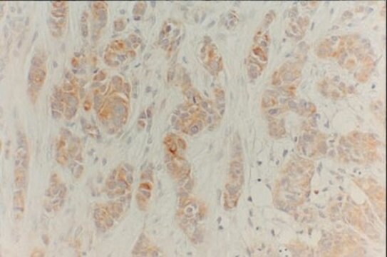
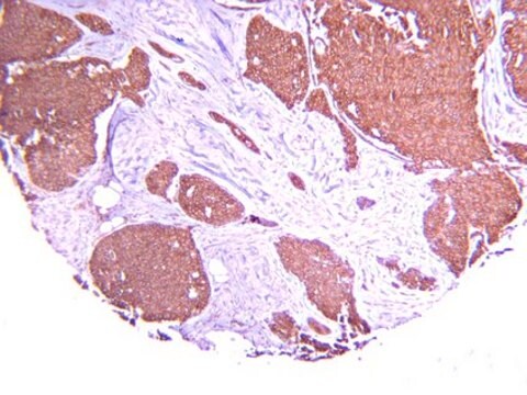
Ki67 antigen is the prototypic cell cycle related nuclear protein, expressed by proliferating cells in all phases of the active cell cycle (G1, S, G2 and M phase). It is absent in resting (G0) cells. Ki67 antibodies are useful in establishing the cell growing fraction in neoplasms (immunohistochemically quantified by determining the number of Ki67 positive cells among the total number of resting cells = Ki67 index). In neoplastic tissues the prognostic value is comparable to the tritiated thymidine labelling index. The correlation between low Ki67 index and histologically low grade tumours is strong. Ki67 is routinely used as a marker of cell cycling and proliferation. This specificity of Ki-S5 antibody for proliferating cells might make it a useful tool for determination of the proliferative fraction in tumors such as Non-Hodgkin′s lymphoma (Kreipe et al., 1993), mammary carcinomas (Mauri et al., 1984), ovarian tumors (Ballin et al., 1994) and prostate cancer.
특이성
In Immunoprecipitation and Western blot experiments the Ki-S5 antibody recognizes a cell-cycle-associated protein of 345 kD and 395 kD (Kreipe et al., 1993) identical with the Ki-67 antigen. The antibody binds to a formalin-resistant epitope of the Ki-67 antigen. The immunoreactivity of Ki-S5 is confined to the nuclei of proliferating cells and no crossreactivity with cytoplasmic antigens of epithelial occurs even after antigen retrieval (Mauri et al., 1984). A comparison of immunohistochemical labeling of fresh and fixed tissue samples of NHL showed that identical results were obtained with Ki-67 and Ki-S5 (Kreipe et al., 1993). The Ki-67 antigen is preferentially expressed during late G1, S, G2 and M phase of the cell cycle, while resting, non-cycling cells (G0 phase) lack the Ki-67 antigen. In addition, constantly proliferating cells (e.g. cell lines) react positively to Ki-S5 during the entire cell-cycle. This specificity of Ki-S5 antibody for proliferating cells might make it a useful tool for determination of the proliferative fraction in tumors such as Non-Hodgkin′s lymphoma (Kreipe et al., 1993), mammary carcinomas (Mauri et al., 1984), ovarian tumors (Ballin et al., 1994) and prostate cancer.
면역원
Nuclear protein preparation from the human cell line U937.50.
애플리케이션
Anti-Ki-67 Antibody, clone Ki-S5 is a high quality Mouse Monoclonal Antibody for the detection of Ki-67 & has been validated in FC, ICC, IHC, IHC(P) & WB.
Immunocytochemistry:
A 5-10 μg/mL concentration of a previous lot was used in IC.
Immunohistochemistry:
A 5-10 μg/mL concentration of a previous lot was used in IH.
Immunohistochemistry:
5-10 µg/mL on formalin fixed paraffin tissue. Citrate buffer-microwave antigen retrieval
Flow Cytometry:
A previous lot of this antibody was used in FC.
Western blot:
1-10 µg/mL
Optimal working dilutions must be determined by end user.
Research Category
Epigenetics & Nuclear Function
Epigenetics & Nuclear Function
Research Sub Category
Cell Cycle, DNA Replication & Repair
Cell Cycle, DNA Replication & Repair
품질
Evaluated by Western Blot on A431 lysates.
Western Blot Analysis:
1:500 dilution of this antibody detected cell-cycle-associated protein of 345 kDa and 395 kDa on 10 μg of A431 lysates.
Western Blot Analysis:
1:500 dilution of this antibody detected cell-cycle-associated protein of 345 kDa and 395 kDa on 10 μg of A431 lysates.
표적 설명
345 & 395 kDa
물리적 형태
Format: Purified
Mouse monoclonal IgG1 in buffer containing 0.02 M Phosphate buffer, 0.25 M NaCl, pH 7.6 with 0.1% sodium azide.
Protein A purified
저장 및 안정성
Stable for 1 year at 2-8ºC from date of receipt.
분석 메모
Control
Tonsil tissue, A431 cell lysate
Tonsil tissue, A431 cell lysate
기타 정보
Concentration: Please refer to the Certificate of Analysis for the lot-specific concentration.
법적 정보
CHEMICON is a registered trademark of Merck KGaA, Darmstadt, Germany
면책조항
Unless otherwise stated in our catalog or other company documentation accompanying the product(s), our products are intended for research use only and are not to be used for any other purpose, which includes but is not limited to, unauthorized commercial uses, in vitro diagnostic uses, ex vivo or in vivo therapeutic uses or any type of consumption or application to humans or animals.
적합한 제품을 찾을 수 없으신가요?
당사의 제품 선택기 도구.을(를) 시도해 보세요.
Storage Class Code
10 - Combustible liquids
WGK
WGK 2
Flash Point (°F)
Not applicable
Flash Point (°C)
Not applicable
시험 성적서(COA)
제품의 로트/배치 번호를 입력하여 시험 성적서(COA)을 검색하십시오. 로트 및 배치 번호는 제품 라벨에 있는 ‘로트’ 또는 ‘배치’라는 용어 뒤에서 찾을 수 있습니다.
이미 열람한 고객
Retinoid-induced epidermal hyperplasia is mediated by epidermal growth factor receptor activation via specific induction of its ligands heparin-binding EGF and amphiregulin in human skin in vivo.
Rittie, L; Varani, J; Kang, S; Voorhees, JJ; Fisher, GJ
The Journal of Investigative Dermatology null
Paula Rocktäschel et al.
Epilepsy & behavior : E&B, 101(Pt B), 106581-106581 (2019-11-26)
Tuberous sclerosis complex (TSC) is a neurodevelopmental disorder caused by deletions in the TSC1 or TSC2 genes that is associated with epilepsy in up to 90% of patients. Seizures are suggested to start in benign brain tumors, cortical tubers, or
Katarzyna Biadasiewicz et al.
Biology of reproduction, 90(5), 101-101 (2014-04-04)
ADAM12, consisting of a membrane-bound (ADAM12L) and a secreted (ADAM12S) form, is expressed exclusively in regenerating and developing tissue as well as in certain cancer types. Strong ADAM12 expression levels have been noticed in the human placenta, and deregulated ADAM12S
H Kreipe et al.
The American journal of pathology, 142(1), 3-9 (1993-01-01)
A monoclonal antibody (Ki-S1) has been raised that reacts with the nuclei of proliferating cells. The antigen recognized is resistant to formalin fixation and can be detected in frozen tissues as well as in routinely processed specimens. In immunohistochemistry, nuclear
Identification of a Src tyrosine kinase/SIAH2 E3 ubiquitin ligase pathway that regulates C/EBP? expression and contributes to transformation of breast tumor cells.
Sarkar, TR; Sharan, S; Wang, J; Pawar, SA; Cantwell, CA; Johnson, PF; Morrison, DK; Wang et al.
Molecular and cellular biology null
자사의 과학자팀은 생명 과학, 재료 과학, 화학 합성, 크로마토그래피, 분석 및 기타 많은 영역을 포함한 모든 과학 분야에 경험이 있습니다..
고객지원팀으로 연락바랍니다.