HPA042257
Anti-RP1 antibody produced in rabbit
Prestige Antibodies® Powered by Atlas Antibodies, affinity isolated antibody, buffered aqueous glycerol solution
別名:
MAPRE2, Microtubule-associated protein RP/EB family member 2, Anti-Dcdc4a, Anti-Retinitis pigmentosa 1 (autosomal dominant)
ログイン組織・契約価格を表示する
すべての画像(3)
About This Item
おすすめの製品
由来生物
rabbit
品質水準
結合体
unconjugated
抗体製品の状態
affinity isolated antibody
抗体製品タイプ
primary antibodies
クローン
polyclonal
製品種目
Prestige Antibodies® Powered by Atlas Antibodies
フォーム
buffered aqueous glycerol solution
交差性
human
テクニック
immunohistochemistry: 1:500-1:1000
免疫原配列
VTCSPCEMCTVNKAYSPKETCNPSDTFFPSDGYGVDQTSMNKACFLGEVCSLTDTVFSDKACAQKENHTYEGACPIDETYVPVNVCNTIDFLNSKENTYTDNLDSTEELERGDDIQKDLNILTDPEYKNGFNTLVSHQNVSNLSSCG
UniProtアクセッション番号
輸送温度
wet ice
保管温度
−20°C
ターゲットの翻訳後修飾
unmodified
遺伝子情報
human ... RP1(6101)
詳細
Retinitis pigmentosa-1 (RP1) is a photoreceptor microtubule-associated protein. RP1 belongs to end binding protein 1(EB1) protein family, which interacts with adenomatous polyposis coli (APC). RP1 is located on human chromosome 8.
免疫原
retinitis pigmentosa 1 (autosomal dominant) recombinant protein epitope signature tag (PrEST)
アプリケーション
All Prestige Antibodies Powered by Atlas Antibodies are developed and validated by the Human Protein Atlas (HPA) project and as a result, are supported by the most extensive characterization in the industry.
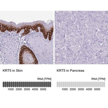
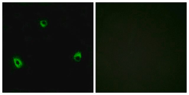
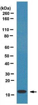
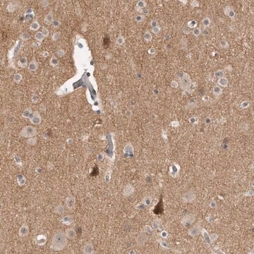
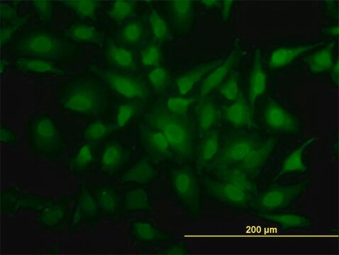
The Human Protein Atlas project can be subdivided into three efforts: Human Tissue Atlas, Cancer Atlas, and Human Cell Atlas. The antibodies that have been generated in support of the Tissue and Cancer Atlas projects have been tested by immunohistochemistry against hundreds of normal and disease tissues and through the recent efforts of the Human Cell Atlas project, many have been characterized by immunofluorescence to map the human proteome not only at the tissue level but now at the subcellular level. These images and the collection of this vast data set can be viewed on the Human Protein Atlas (HPA) site by clicking on the Image Gallery link. We also provide Prestige Antibodies® protocols and other useful information.
The Human Protein Atlas project can be subdivided into three efforts: Human Tissue Atlas, Cancer Atlas, and Human Cell Atlas. The antibodies that have been generated in support of the Tissue and Cancer Atlas projects have been tested by immunohistochemistry against hundreds of normal and disease tissues and through the recent efforts of the Human Cell Atlas project, many have been characterized by immunofluorescence to map the human proteome not only at the tissue level but now at the subcellular level. These images and the collection of this vast data set can be viewed on the Human Protein Atlas (HPA) site by clicking on the Image Gallery link. We also provide Prestige Antibodies® protocols and other useful information.
生物化学的/生理学的作用
Retinitis pigmentosa-1 (RP1) regulates microtubule formation. Casein kinase II (CK2) a protein kinase when phosphorylates RP1 at Ser(236) aids cell adhesion.RP1 contributes to maintain the architecture of photoreceptor outer segments. Dysfunctioning of RP1 leads to neurodegenerative diseases. Mutations in RP1 gene is known to cause retinitis pigmentosa. Certain regions of RP1 may also cause autosomal recessive rod-cone dystrophy.
特徴および利点
Prestige Antibodies® are highly characterized and extensively validated antibodies with the added benefit of all available characterization data for each target being accessible via the Human Protein Atlas portal linked just below the product name at the top of this page. The uniqueness and low cross-reactivity of the Prestige Antibodies® to other proteins are due to a thorough selection of antigen regions, affinity purification, and stringent selection. Prestige antigen controls are available for every corresponding Prestige Antibody and can be found in the linkage section.
Every Prestige Antibody is tested in the following ways:
Every Prestige Antibody is tested in the following ways:
- IHC tissue array of 44 normal human tissues and 20 of the most common cancer type tissues.
- Protein array of 364 human recombinant protein fragments.
関連事項
Corresponding Antigen APREST71374
物理的形状
PBS溶液(pH 7.2、40%グリセロール、0.02% アジ化ナトリウム含有)
法的情報
Prestige Antibodies is a registered trademark of Merck KGaA, Darmstadt, Germany
適切な製品が見つかりませんか。
製品選択ツール.をお試しください
保管分類コード
10 - Combustible liquids
WGK
WGK 1
引火点(°F)
Not applicable
引火点(℃)
Not applicable
最新バージョンのいずれかを選択してください:
Targeted next generation sequencing identifies novel mutations in RP1 as a relatively common cause of autosomal recessive rod-cone dystrophy
El Shamie, et al.
BioMed Research International, 2015 (2015)
The retinitis pigmentosa 1 protein is a photoreceptor microtubule-associated protein
Liu, Qin, et al.
The Journal of Neuroscience, 24(29), 6427-6436 (2004)
Linkage mapping of autosomal dominant retinitis pigmentosa (RP1) to the pericentric region of human chromosome 8
Blanton,, et al.
Genomics, 11(4), 857-869 (1991)
RP1 is a phosphorylation target of CK2 and is involved in cell adhesion
Stenner,, et al.
PLoS ONE, 8(7), e67595-e67595 (2013)
Molecular basis for photoreceptor outer segment architecture
Goldberg,, et al.
Progress in Retinal and Eye Research, 55, 52-81 (2016)
Global Trade Item Number
| カタログ番号 | GTIN |
|---|---|
| HPA042257-25UL | 4061841180050 |
| HPA042257-100UL | 4061835737116 |
ライフサイエンス、有機合成、材料科学、クロマトグラフィー、分析など、あらゆる分野の研究に経験のあるメンバーがおります。.
製品に関するお問い合わせはこちら(テクニカルサービス)