
おすすめの製品


由来生物
rabbit
結合体
unconjugated
抗体製品の状態
affinity isolated antibody
抗体製品タイプ
primary antibodies
クローン
polyclonal
製品種目
Prestige Antibodies® Powered by Atlas Antibodies
フォーム
buffered aqueous glycerol solution
交差性
human
テクニック
immunohistochemistry: 1:50- 1:200
免疫原配列
DEMYKILLNDYEYRQKQILMENAELKKVLQQMKKEMISLLSPQKKKPRERVDDSTGTVISDVEEDAGELSRESMWDLSCETVREQLTNSIRKQWRILKSHVEKLDNQVSKVHLEGFNDEDVISRQDHEQETE
UniProtアクセッション番号
輸送温度
wet ice
保管温度
−20°C
ターゲットの翻訳後修飾
unmodified
遺伝子情報
human ... SSX2IP(117178)
免疫原
アファディン・α-アクチニン結合タンパク質のPrEST(protein epitope signature tag)抗原リコンビナントタンパク質。
アプリケーション
Prestige抗体®は、Human Proteome Resource(HPR)プロジェクト(www.proteinatlas.org)によって開発・実証されたAtlas抗体です。抗体はすべて、数百の正常組織・疾病組織に対する免疫組織染色試験を行っています。これらの染色画像はHuman Protein Atlas(HPA)サイトで[Image Gallery]リンクをクリックするとご覧いただけます。ほとんどのPrestige抗体はプロテインアレイおよびウェスタンブロッティングの試験を行っています。試験のプロトコールおよびPrestige抗体、HPAに関する情報はsigma.com/prestigeをご覧ください。
特徴および利点
Prestige Antibodies® are highly characterized and extensively validated antibodies with the added benefit of all available characterization data for each target being accessible via the Human Protein Atlas portal linked just below the product name at the top of this page. The uniqueness and low cross-reactivity of the Prestige Antibodies® to other proteins are due to a thorough selection of antigen regions, affinity purification, and stringent selection. Prestige antigen controls are available for every corresponding Prestige Antibody and can be found in the linkage section.
Every Prestige Antibody is tested in the following ways:
Every Prestige Antibody is tested in the following ways:
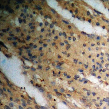
- IHC tissue array of 44 normal human tissues and 20 of the most common cancer type tissues.
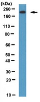
- Protein array of 364 human recombinant protein fragments.
関連事項
Corresponding Antigen APREST70081
物理的形状
PBS溶液(pH 7.2, 40%グリセロールおよび0.02%アジ化ナトリウム含有)。
法的情報
Prestige Antibodies is a registered trademark of Merck KGaA, Darmstadt, Germany
免責事項
Unless otherwise stated in our catalog or other company documentation accompanying the product(s), our products are intended for research use only and are not to be used for any other purpose, which includes but is not limited to, unauthorized commercial uses, in vitro diagnostic uses, ex vivo or in vivo therapeutic uses or any type of consumption or application to humans or animals.
適切な製品が見つかりませんか。
製品選択ツール.をお試しください
保管分類コード
10 - Combustible liquids
WGK
WGK 1
引火点(°F)
Not applicable
引火点(℃)
Not applicable
適用法令
試験研究用途を考慮した関連法令を主に挙げております。化学物質以外については、一部の情報のみ提供しています。 製品を安全かつ合法的に使用することは、使用者の義務です。最新情報により修正される場合があります。WEBの反映には時間を要することがあるため、適宜SDSをご参照ください。
Jan Code
HPA027306-100UL:
HPA027306-25UL:
最新バージョンのいずれかを選択してください:
Akiko Hori et al.
EMBO reports, 17(3), 326-337 (2016-01-13)
Centrioles are the major constituents of the animal centrosome, in which Plk4 kinase serves as a master regulator of the duplication cycle. Many eukaryotes also contain numerous peripheral particles known as centriolar satellites. While centriolar satellites aid centriole assembly and
Akiko Hori et al.
Biochemical and biophysical research communications, 468(1-2), 39-45 (2015-11-08)
The centrosome plays a pivotal role in a wide range of cellular processes and its dysfunction is causally linked to many human diseases including cancer and developmental and neurological disorders. This organelle contains more than one hundred components, and yet
Maxim A X Tollenaere et al.
Nature communications, 6, 10075-10075 (2015-12-01)
Centriolar satellites (CS) are small granular structures that cluster in the vicinity of centrosomes. CS are highly susceptible to stress stimuli, triggering abrupt displacement of key CS factors. Here we discover a linear p38-MK2-14-3-3 signalling pathway that specifically targets CEP131
Julie C Nielsen et al.
Cells, 7(7) (2018-06-23)
Centriolar satellites (CS) are small proteinaceous granules that cluster around the centrosome and serve as cargo vehicles for centrosomal proteins. It is generally accepted that CS support a number of canonical and specialized centrosome functions. Consequently, these highly dynamic structures
Chun So et al.
Science (New York, N.Y.), 364(6447) (2019-06-30)
Mammalian oocytes segregate chromosomes with a microtubule spindle that lacks centrosomes, but the mechanisms by which acentrosomal spindles are organized and function are largely unclear. In this study, we identify a conserved subcellular structure in mammalian oocytes that forms by
ライフサイエンス、有機合成、材料科学、クロマトグラフィー、分析など、あらゆる分野の研究に経験のあるメンバーがおります。.
製品に関するお問い合わせはこちら(テクニカルサービス)