
おすすめの製品


メーカー/製品名
Roche
包装
kit of for 50 isolations
詳細
Low to medium throughput isolation of total RNA from free FFPE tissue sections in micro scale.
アプリケーション
The High Pure FFPE RNA Micro Kit isolates total RNA from formalin-fixed, paraffin-embedded (FFPE) tissue samples for direct use in:
- Qualitative RT-PCR
- Relative quantification of mRNA with real-time PCR systems such as the LightCycler® 480 System
- Differential display RT-PCR
- cDNA synthesis
- Primer extension
特徴および利点
Nucleic acids bind to the surface of the glass fiber fleece in the presence of a chaotropic salt (guanidine HCl). This allows the High Pure filter tube to specifically immobilize nucleic acids (both DNA and RNA) while they are freed of contaminants. For RNA isolation, the binding conditions can be optimized to ensure immobilization of all the RNA.
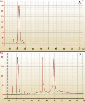
Size Distribution: The typical size of RNA isolated from formalin-fixed tissue ranges from 150 to 1500 bases. However, section thickness, tissue type, age of sample, and the fixation protocol used can affect the yield and quality of the isolated RNA.
Capacity: The High Pure Micro Filter Tubes hold up to 500 μl sample volume.
Sample Material: 1 - 10 μm sections from formalin-fixed, paraffin-embedded (FFPE) tissue (e.g., from colon, breast, liver, kidney, spleen of mammalian species).
The High Pure FFPE RNA Micro Kit isolates total RNA from formalin-fixed, paraffin-embedded (FFPE) tissue samples for direct use in RT-PCR.
- Streamline and simplify RNA isolation (even small RNA fragments) from FFPE tissue.
- Obtain a highly concentrated, ready-to-use eluate and excellent recovery of RNA (>80%).
- Isolate DNA-free RNA for use in qualitative and quantitative RT-PCR.
- Minimize RNA loss with a kit that removes contaminants without precipitation or other handling steps that degrade RNA.
- Generate high-quality template RNA that shows excellent performance and linearity in RT-PCR.
構成
- Tissue Lysis Buffer
- Proteinase K, recombinant, PCR Grade
- Binding Buffer
- Wash Buffer I
- Wash Buffer II
- DNase I, recombinant, lyophilized
- DNase Incubation Buffer
- Elution Buffer
- High Pure Micro Filter Tubes
- Collection Tubes
品質
Formalin-fixed, paraffin-embedded tissue sections are homogenized by overnight Proteinase K digestion and purified as described. RNA yield is determined by measuring the optical density at 260 nm.
The RNA eluate and specific primers for the ß2M gene are used in one-step RT-PCR. In the following PCR on the LightCycler® 2.0 Instrument (accomplished using the LightCycler® RNA Amplification Kit SYBR Green I and specific primers for β2M), the expected amplification signal is obtained at a Cp-value less than 24.
Absence of contaminating genomic DNA is examined by PCR on a LightCycler® 2.0 Instrument without a reverse transcriptase step; no amplification product is obtained.
調製ノート
To prepare tissue sections for RNA isolation, fixation reagents must be removed from the samples; after deparaffinization, the sections are ready to be processed with the High Pure FFPE RNA Micro Kit. Deparaffinized tissue samples are disrupted and homogenized during incubation with Proteinase K and a chaotropic salt (guanidine HCl). The homogenate is then applied to the glass fiber fleece in a High Pure Micro Filter Tube.
Under the buffer conditions used in the procedure, all nucleic acids bind specifically to the glass fiber fleece, while contaminating substances (salts, proteins, and other tissue contaminants) do not. DNA in the preparation is digested with DNase I directly on the filter. Brief wash-and-spin steps readily remove the digested DNA fragments and other contaminating substances. The remaining purified RNA is then eluted in a small volume of low-salt buffer.
Under the buffer conditions used in the procedure, all nucleic acids bind specifically to the glass fiber fleece, while contaminating substances (salts, proteins, and other tissue contaminants) do not. DNA in the preparation is digested with DNase I directly on the filter. Brief wash-and-spin steps readily remove the digested DNA fragments and other contaminating substances. The remaining purified RNA is then eluted in a small volume of low-salt buffer.
アナリシスノート
Typical RNA Recovery
Starting Material and Quantity: 1 - 10 μm FFPE sections, colon, breast, liver, kidney, spleen of mammalian species
Yield/Recovery: 1.5 - 3.5 μg/5 μm section
Time Required: 60 minutes without 3 hour incubation
Number of Reactions: 50/1-10 μm sections
Starting Material and Quantity: 1 - 10 μm FFPE sections, colon, breast, liver, kidney, spleen of mammalian species
Yield/Recovery: 1.5 - 3.5 μg/5 μm section
Time Required: 60 minutes without 3 hour incubation
Number of Reactions: 50/1-10 μm sections
法的情報
LightCycler is a registered trademark of Roche


シグナルワード
Danger
危険有害性の分類
Acute Tox. 4 Dermal - Acute Tox. 4 Inhalation - Acute Tox. 4 Oral - Aquatic Chronic 3 - Eye Dam. 1 - Resp. Sens. 1 - Skin Corr. 1C - Skin Sens. 1 - STOT SE 3
ターゲットの組織
Respiratory system
補足的ハザード
保管分類コード
13 - Non Combustible Solids
WGK
WGK 2
引火点(°F)
does not flash
引火点(℃)
does not flash
この製品を見ている人はこちらもチェック
Zachary Kerr et al.
Head and neck pathology, 14(3), 577-587 (2019-09-14)
Kallikrein-related peptidases (KLKs) are a group of 15 serine proteases implicated in a variety of biological processes. Aberrant expression of KLKs has been associated with the development of certain cancers. However, the role of KLKs in salivary tumors has not
Isabella Wimmer et al.
Scientific reports, 8(1), 6351-6351 (2018-04-22)
Formalin-fixed paraffin-embedded (FFPE) tissues are valuable resources commonly used in pathology. However, formalin fixation modifies nucleic acids challenging the isolation of high-quality RNA for genetic profiling. Here, we assessed feasibility and reliability of microarray studies analysing transcriptome data from fresh
David S Shames et al.
Clinical cancer research : an official journal of the American Association for Cancer Research, 19(24), 6912-6923 (2013-10-08)
We sought to identify predictive biomarkers for a novel nicotinamide phosphoribosyltransferase (NAMPT) inhibitor. We use a NAMPT inhibitor, GNE-617, to evaluate nicotinic acid rescue status in a panel of more than 400 cancer cell lines. Using correlative analysis and RNA
Anna R Tröscher et al.
Acta neuropathologica, 137(4), 619-635 (2019-01-22)
Microglia nodule formation is a common feature in inflammatory brain diseases mediated by T lymphocytes such as viral and paraneoplastic encephalitis, multiple sclerosis, and Rasmussen encephalitis (RE). However, its role has not been fully understood yet. We hypothesized that, in
Jun Liu et al.
Nature cell biology, 20(9), 1074-1083 (2018-08-30)
N6-methyladenosine (m6A) messenger RNA methylation is a gene regulatory mechanism affecting cell differentiation and proliferation in development and cancer. To study the roles of m6A mRNA methylation in cell proliferation and tumorigenicity, we investigated human endometrial cancer in which a
ライフサイエンス、有機合成、材料科学、クロマトグラフィー、分析など、あらゆる分野の研究に経験のあるメンバーがおります。.
製品に関するお問い合わせはこちら(テクニカルサービス)