
PICM0RG50
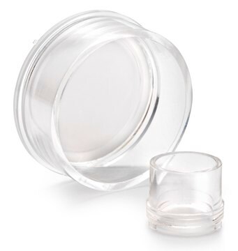
Millicell細胞培養インサート 30 mm 親水性PTFE 0.4 µm
pore size 0.4 μm, diam. 30 mm, transparent PTFE membrane, hydrophilic, H 5 mm, size 6 wells, sterile
別名:
Millicell Cell Culture Insert, 30 mm, hydrophilic PTFE, 0.4 µm, Millicell-CM, cell culture inserts, organotypic inserts, plate inserts
About This Item
おすすめの製品

物質
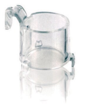
polystyrene housing
transparent PTFE membrane
品質水準
無菌性
ethylene oxide treated
sterile
特徴
hydrophilic
メーカー/製品名
Millicell®
包装
pkg of Individually blister packaged
パラメーター
50 °C max. temp.
テクニック
cell attachment: suitable
cell culture | mammalian: suitable
cell differentiation: suitable
H
5 mm
直径
30 mm
サイズ
6 wells
表面積
4.2 cm2
色
transparent membrane, when wetted
Matrix
Biopore™
ポアサイズ
0.4 μm
結合型
low binding surface
検出方法
fluorometric
輸送温度
ambient
詳細
アプリケーション
- 細胞接着、3D細胞培養、細胞増殖、細胞分化、免疫細胞染色
- Bioporeメンブレン(親水性PTFE)は、低タンパク質結合、生細胞観察、免疫蛍光の用途に最適です。
特徴および利点
- 高い細胞生存率と3D外植片構造の優れた研究用
- 高さが低いため、標準的なシャーレの中に収まります
- Biopore(PTFE)メンブレンは、40日にもわたる高い生存率と、優れた膜透過酸素輸送を実現します——
- メンブレンは、光学的に透明であり、長期的な器官型外植片維持に最適化されています
調製ノート
- Add assay solution or media to the receiver well: 2 mL for 6 well format orequivalent dimensions.
- With forceps, place the insert into the receiver well at a 30°-45° angle togradually wet it out from the basolateral side.
- Once edges start to wet out, gently lower the insert until it is fully seatedinto the well.
法的情報
保管分類コード
10-13 - German Storage Class 10 to 13
試験成績書(COA)
製品のロット番号・バッチ番号を入力して、試験成績書(COA) を検索できます。ロット番号・バッチ番号は、製品ラベルに「Lot」または「Batch」に続いて記載されています。
この製品を見ている人はこちらもチェック


プロトコル
This is a Toluidine Blue Staining protocol.
This is a Toluidine Blue Staining protocol listing materials and methods.
This protocol covers 3 modes for the microscopic examination of cell samples.
This protocol covers 3 modes for the microscopic examination of cell samples.
関連コンテンツ
This page covers the ECM coating protocols developed for four types of ECMs on Millicell®-CM inserts, Collagen Type 1, Fibronectin, Laminin, and Matrigel.
This page covers the ECM coating protocols developed for four types of ECMs on Millicell®-CM inserts, Collagen Type 1, Fibronectin, Laminin, and Matrigel.
ライフサイエンス、有機合成、材料科学、クロマトグラフィー、分析など、あらゆる分野の研究に経験のあるメンバーがおります。.
製品に関するお問い合わせはこちら(テクニカルサービス)